Category:Microscopic images of actin stained with phalloidin
Jump to navigation
Jump to search
Subcategories
This category has only the following subcategory.
Media in category "Microscopic images of actin stained with phalloidin"
The following 48 files are in this category, out of 48 total.
-
3D-SIM-4 Anaphase 3 color.jpg 954 × 724; 264 KB
-
Actin-cortex.png 1,500 × 686; 579 KB
-
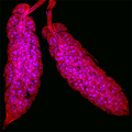
Aedes aegypti adult ovaries.png 656 × 656; 401 KB
-
Antiguas(Cell migrating)(RGB).tif 799 × 775; 1.79 MB
-
BioTek-Wikipedia-Image.tif 852 × 630; 2.6 MB
-
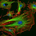
Bovine Pulmonary Artery Endothelial Cells Fluorescent Image 2.jpg 1,920 × 1,452; 298 KB
-
Bovine Pulmonary Artery Endothelial Cells Fluorescent Image 3.jpg 1,920 × 1,452; 423 KB
-
Bovine Pulmonary Artery Endothelial Cells Fluorescent Image.jpg 1,920 × 1,452; 304 KB
-
Cannabioid agonist WIN acting on pancreatic stellate cell.png 2,000 × 2,516; 1.85 MB
-
Cell division.tif 1,615 × 1,357, 3 pages; 8.42 MB
-
Confocal micrograph of stained HeLa.jpg 1,345 × 1,345; 883 KB
-
Cultured schwann cell.jpg 151 × 443; 22 KB
-
Depth Coded Phalloidin Stained Actin Filaments Cancer Cell.png 3,176 × 2,472; 9.86 MB
-
Fibroblast,polydopamine nanoparticles and F-actin.jpg 2,048 × 2,048; 634 KB
-
FluorescentCells.jpg 512 × 512; 56 KB
-
HEK 293.jpg 4,785 × 4,620; 10.51 MB
-
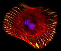
HELA cells stained with DAPI and Phalloidin.tiff 1,376 × 1,038; 4.1 MB
-
HeLa-II.jpg 3,200 × 2,400; 2.76 MB
-
Immunology seen from the sky.png 2,048 × 2,048; 10.26 MB
-
Indian Muntjac fibroblast cells (23725924864).jpg 1,924 × 1,218; 718 KB
-
Indian Muntjac fibroblast cells (24271618921).jpg 1,923 × 1,210; 684 KB
-
Indian Muntjac fibroblast cells (24327908636).jpg 1,920 × 1,217; 1,007 KB
-
MAX 052913 STED Phallloidin.png 4,089 × 4,093; 8.94 MB
-
MEF microfilaments.jpg 600 × 450; 58 KB
-
Morphology-and-morphometry-of-the-hepatopancreas-of-O.jpg 744 × 535; 151 KB
-
Mouse Intestine (23727287703).jpg 1,915 × 1,217; 1,010 KB
-
Mouse Kidney (23725924684).jpg 1,916 × 1,210; 993 KB
-
MultiPhotonExcitation-Fig10-doi10.1186slash1475-925X-5-36.JPEG 2,133 × 2,133; 949 KB
-
Myoanatomical changes in Richtersius coronifer during tun formation.png 2,049 × 2,615; 3.88 MB
-
Nanotubes.png 2,128 × 1,632; 2.29 MB
-
Nesprin2InCos7Zellen.jpg 486 × 486; 48 KB
-
Pancreatic stellate cell cropped.png 1,001 × 1,570; 769 KB
-
PC3 prostate cancer cells, confocal image, 63x.jpg 1,472 × 1,474; 304 KB
-
Phalloidin staining of actin filaments.tif 4,008 × 3,275; 9.59 MB
-
Polyps of Cnidaria colony.jpg 5,184 × 3,456; 546 KB
-
Single polyp of Gonothyraea loveni.jpg 3,456 × 5,184; 936 KB
-
Sphaeroforma Arctica (28638788957).jpg 564 × 564; 38 KB
-
STD Depth Coded Stack Phallodin Stained Actin Filaments.png 4,096 × 4,096; 21.54 MB
-
STD Depth Coded Stack Slices through Cells.png 5,000 × 3,893; 22.96 MB
-
STED Confocal Comparison 50nm HWFM.png 5,399 × 4,263; 10.59 MB
-
Striped elegance.jpg 1,024 × 1,024; 201 KB
-
SUM 110913 Cort Neurons 2.5d in vitro 488 Phalloidin no perm 4 cmle-2.png 4,096 × 2,584; 4.33 MB
-
The kinocilia is mislocalized in Tailchaser hair cells.png 2,989 × 2,746; 6.19 MB
-
The Mediterranean Saint-Pierre.pdf 29,941 × 29,941; 15.26 MB
-
Treasure Island.png 2,048 × 2,048; 2.31 MB


