Category:Metaphase
Jump to navigation
Jump to search
cell cycle phase, following prophase or prometaphase in higher eukaryotes, during which chromosomes become aligned on the equatorial plate of the cell | |||||
| Upload media | |||||
| Instance of | |||||
|---|---|---|---|---|---|
| Subclass of |
| ||||
| Part of |
| ||||
| Follows | |||||
| Followed by | |||||
| |||||
Subcategories
This category has only the following subcategory.
M
Media in category "Metaphase"
The following 63 files are in this category, out of 63 total.
-
Allium-Mitose03-DM100x BL28.jpg 859 × 942; 420 KB
-
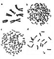
Bcrablmet.jpg 213 × 244; 10 KB
-
Chicken microchromosomes.tif 497 × 478; 720 KB
-
ChickenChromosomesBMC Genomics5-56Fig4.jpg 827 × 955; 40 KB
-
Chr2 orang human.jpg 484 × 275; 42 KB
-
Chromatin fibers (261 19) Mitosis; Xenopus egg.jpg 3,751 × 2,401; 2.24 MB
-
Chromosome Attachement.jpg 836 × 464; 40 KB
-
Chromosome-Dynamics-Visualized-with-an-Anti-Centromeric-Histone-H3-Antibody-in-Allium-pone.0051315.s006.ogv 8.0 s, 1,280 × 720; 2.12 MB
-
Cytogenetic Mega-telomeres GGA 9 and W.jpg 488 × 441; 35 KB
-
De-Metaphase.ogg 1.9 s; 19 KB
-
Drosophila metaphase chromosomes.jpg 950 × 508; 25 KB
-
Examples of mammalian chromosomes.jpeg 797 × 869; 67 KB
-
Fuseau mitotique mitosis.jpg 977 × 591; 68 KB
-
Human female metaphase chromosomes.tif 659 × 647; 1.24 MB
-
HumanChromosomesChromomycinA3.jpg 453 × 438; 29 KB
-
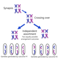
Independent assortment.png 2,287 × 2,186; 558 KB
-
Kinetochore.jpg 400 × 317; 136 KB
-
Meiosis (263 08).jpg 3,749 × 2,399; 918 KB
-
Metaphase (261 04) Pressed; meristem of root; onion.jpg 1,600 × 1,024; 339 KB
-
Metaphase (261 05) Pressed; meristem of root; onion.jpg 3,751 × 2,401; 1.41 MB
-
Metaphase (261 06) Pressed; meristem of root; onion.jpg 3,751 × 2,401; 1.34 MB
-
Metaphase (261 21) Pressed; meristem of root; onion.jpg 3,751 × 2,401; 1.79 MB
-
Metaphase (261 22) Pressed; meristem of root; onion.jpg 3,751 × 2,401; 1.75 MB
-
Metaphase chromosomes.jpg 399 × 192; 55 KB
-
Metaphase during Mitosis.svg 512 × 431; 49 KB
-
Metaphase eukaryotic mitosis.svg 994 × 699; 125 KB
-
Metaphase Spread of Rat.jpg 3,072 × 2,304; 1.16 MB
-
Metaphase spread of the Siberian Roe deer (Capreolus pygargus).jpg 391 × 452; 26 KB
-
Metaphase spread of the Viscacha rat (Tympanoctomys barrerae).jpg 404 × 385; 22 KB
-
Metaphase-plate-of-aspen-clone-Meshabash--2n38.jpg 660 × 536; 147 KB
-
Metaphase.jpg 160 × 160; 23 KB
-
Metaphase.png 394 × 301; 31 KB
-
Metaphase1.jpg 4,032 × 3,024; 1.43 MB
-
Metaphase3.jpg 3,024 × 4,032; 1.49 MB
-
Metaphase5.jpg 626 × 470; 77 KB
-
Metaphase6.jpg 1,280 × 960; 122 KB
-
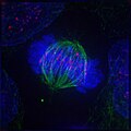
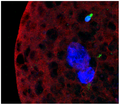
MetaphaseIF.jpg 384 × 384; 119 KB
-
Mitotic Metaphase.svg 97 × 81; 78 KB
-
Prometaphase in a crane-fly spermatocyte treated with 50 nM Calyculin A - 1475-9268-6-1-S3.mpg.ogv 0.0 s, 312 × 240; 2.41 MB
-
-
-
-
SCE Metaphase-BMC Cell Biol 2-11-6.jpg 899 × 897; 68 KB
-
Schmetaphase.png 644 × 658; 387 KB
-
Schnoymetaphase.png 736 × 334; 31 KB
-
Secondharmbatio3.png 427 × 393; 118 KB
-
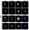
SiCENP-E metaphase.png 501 × 510; 97 KB
-
Stages of early mitosis in a vertebrate cell (hy).svg 512 × 239; 69 KB
-
Stages of early mitosis in a vertebrate cell with micrographs of chromatids.svg 512 × 1,170; 458 KB
-
-
-
-
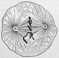
Wilson1900Fig31A.jpg 534 × 525; 83 KB
-
Метафаза-I.jpg 869 × 353; 107 KB
-
Метафаза-II.jpg 902 × 439; 137 KB


