Category:Medical technology
Jump to navigation
Jump to search
health sciences field | |||||
| Upload media | |||||
| Instance of |
| ||||
|---|---|---|---|---|---|
| Subclass of | |||||
| |||||
Subcategories
This category has the following 22 subcategories, out of 22 total.
A
B
C
D
F
- Femtech (6 F)
I
M
- Ludwig Müller-Uri (3 F)
P
- PLANFOX (1 F)
R
- Rapid Transfer Port (9 F)
S
V
- Veronica Bucket (54 F)
Pages in category "Medical technology"
This category contains only the following page.
Media in category "Medical technology"
The following 159 files are in this category, out of 159 total.
-
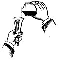
"How to Measure" diagram, with graduated cylinder measuring fluid drams, 1926.jpg 1,648 × 1,674; 449 KB
-
1 (13) resized.jpg 2,458 × 1,635; 583 KB
-
Abaisse-langue pour narcose ( ou écarte-bouche pour narcose) - 480.jpg 2,751 × 2,024; 2.95 MB
-
Absaugpumpe manuell.jpg 800 × 600; 134 KB
-
Afterloading Applicators.jpg 4,000 × 3,000; 4.2 MB
-
Afterloading Device latView.jpg 2,719 × 3,848; 5.03 MB
-
Afterloading schematicDrawing.pdf 1,239 × 1,754; 20 KB
-
AL Geraet frontal.png 1,786 × 3,366; 5.41 MB
-
Analdehner.jpg 618 × 1,110; 37 KB
-
Antonio e Biagio e Cesare Arrigo Cryoconservation room.jpg 2,448 × 3,264; 1.26 MB
-
Aplicación de la tecnología en la medicina.pdf 1,275 × 1,650, 3 pages; 83 KB
-
AVL Logo.svg 886 × 887; 2 KB
-
BBraun Space.jpg 1,542 × 2,028; 131 KB
-
Biotronik logo.svg 1,024 × 137; 7 KB
-
Blasenkatheter.PNG 898 × 429; 10 KB
-
BOWA LOGO.png 945 × 230; 30 KB
-
Boîte de coupes histologiques - IHM-0253.jpg 3,088 × 2,056; 2.76 MB
-
Braun Perfusor.jpg 1,950 × 654; 279 KB
-
C-fos marker (TI Non-invasive DBS).png 430 × 303; 159 KB
-
CAD Modellation der Infix®-Krone.jpg 1,100 × 786; 329 KB
-
CAD Modellation der Infix®-Krone2.jpg 1,100 × 786; 313 KB
-
CAD Modellation Verblendung.tiff 906 × 834; 384 KB
-
CAD-Modellation Gerüst.jpg 769 × 716; 105 KB
-
CAD-Modellation Verblendung.jpg 906 × 834; 39 KB
-
CAD-Modellation Verblendung2.jpg 895 × 834; 39 KB
-
Cliniporator 2 IGEA.JPG 2,304 × 3,072; 4.7 MB
-
Combo bolus.JPG 576 × 364; 35 KB
-
Covalent binding for Wiki project.jpg 486 × 127; 15 KB
-
Darmmodell.JPG 570 × 858; 87 KB
-
Dauerschwingfestigkeit Infix®-Krone.jpg 1,280 × 985; 57 KB
-
De-Medizintechnik.ogg 2.6 s; 26 KB
-
Desfibrilador público.jpg 778 × 159; 130 KB
-
Diagram przeplywu informacji teleoperacje.svg 1,453 × 638; 13 KB
-
Digital verblendete Gerüststruktur.jpg 769 × 716; 116 KB
-
Domo Origato Surgeon Roboto (230350985).jpeg 1,536 × 2,048; 1.04 MB
-
Electrode cgm.jpg 410 × 500; 25 KB
-
ElectronicHealthRecord-Beratungsursache ProDok Screenshot.jpg 484 × 396; 45 KB
-
EMT Emergency & Military Tourniquet.jpg 1,200 × 900; 286 KB
-
ERBE Elektromedizin logo.svg 1,024 × 345; 3 KB
-
ERBE Erbogalvan-Comfort und Erbosonat Comfort.jpg 3,292 × 2,450; 923 KB
-
Estacion de trabajo medical Keosys.JPG 2,048 × 1,536; 700 KB
-
First technetium-99m generator - 1958.jpg 1,392 × 1,788; 1.03 MB
-
Five99mTechnetiumGenerators.jpg 254 × 239; 15 KB
-
Fotothek df roe-neg 0006074 025 Messestand mit medizinischen Geräten.jpg 800 × 549; 115 KB
-
Fotothek df roe-neg 0006490 034 Medizintechnische Geräte.jpg 800 × 542; 143 KB
-
Freestyle.png 1,347 × 1,962; 1.91 MB
-
Gehirnmodell.JPG 600 × 488; 112 KB
-
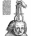
Gersdorff - Schädelwunde.jpg 546 × 646; 99 KB
-
Ghislaine Alajouanine.JPG 533 × 800; 95 KB
-
Glukometr (OneTouch Ultra).jpg 1,000 × 767; 208 KB
-
Handheld medical tube sealer 1.svg 512 × 836; 6 KB
-
Handheld medical tube sealer 2.svg 512 × 274; 8 KB
-
HFOV.jpeg 393 × 705; 118 KB
-
Hickman line.png 304 × 692; 366 KB
-
Hungarian Medical Helicopter.jpg 5,472 × 3,648; 12.26 MB
-
Imaging video taken on a COVID-19 lung ultrasound phantom.webm 3.1 s, 958 × 460; 587 KB
-
Infix®-Brücke as machined2.jpg 1,168 × 886; 204 KB
-
Infix®-Brücke2.jpg 886 × 672; 95 KB
-
Infix®-Krone as machined.jpg 1,168 × 886; 367 KB
-
Infix®-Krone.jpg 1,051 × 886; 127 KB
-
Infix®-Krone2.jpg 1,051 × 797; 122 KB
-
Infusionsbesteck.JPG 1,632 × 1,224; 202 KB
-
Injectomat1.JPG 3,611 × 2,377; 4.51 MB
-
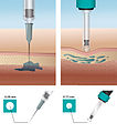
Injektion intradermal.jpg 1,473 × 1,663; 701 KB
-
Insulin pennadel.jpg 3,008 × 2,000; 1.11 MB
-
Insulinpen Helinos 01.jpg 2,816 × 2,112; 1.42 MB
-
Insulinpen Helinos 02.jpg 2,816 × 2,112; 1.43 MB
-
InVivoSemiconductorProbes.jpg 4,000 × 3,000; 4.34 MB
-
IUDCPCopperT380A.gif 269 × 417; 54 KB
-
Kalt-warm-kompresse-gel-mikrowellen-geeignet-1-st-13-x-14-cm 1 1 1.jpg 1,000 × 1,000; 65 KB
-
Kalt-warm-kompresse-gel-mikrowellen-geeignet-1-st. 1 1.jpg 1,000 × 1,000; 62 KB
-
KitSOS spirometre.JPG 494 × 352; 79 KB
-
Koexistenztest 013 klein.jpg 1,063 × 797; 302 KB
-
Labka AAS HP1000 23 aar.jpg 800 × 600; 506 KB
-
Labka brugervejledninger.jpg 3,209 × 2,887; 1.26 MB
-
Labka CSI 101.jpg 5,351 × 1,258; 1,004 KB
-
Labka database.jpg 4,368 × 2,727; 1.5 MB
-
Labka kum 1988 p.jpg 3,484 × 3,875; 2.49 MB
-
Labka kum 2001 p.jpg 1,341 × 1,805; 792 KB
-
Labka LSP opbygning.jpg 3,768 × 2,843; 1.26 MB
-
Labka produktion.jpg 612 × 868; 137 KB
-
Labka reference.jpg 528 × 852; 113 KB
-
Labka rek 2 p.png 804 × 633; 32 KB
-
Labka RH HP1000 21MX.jpg 2,562 × 3,693; 1.34 MB
-
Labka skaermbillede meny.jpg 4,378 × 2,919; 1.7 MB
-
Laka arbejdsregister.jpg 5,472 × 3,648; 3.51 MB
-
Logo Mathys.jpg 176 × 57; 11 KB
-
Loops male normal and ischemic.jpg 1,367 × 643; 118 KB
-
LSP brugere.jpg 813 × 581; 73 KB
-
LSP ptb p.jpg 1,339 × 1,869; 873 KB
-
LSP rek 1 p.png 811 × 612; 36 KB
-
LSP søg 1 p.png 815 × 634; 29 KB
-
LSP søg 2 p.jpg 817 × 612; 178 KB
-
LSP søg 2.jpg 817 × 612; 178 KB
-
MathysEuropean CL.png 426 × 137; 12 KB
-
Measurement2.gif 677 × 276; 28 KB
-
Measurement3.gif 667 × 218; 22 KB
-
Medical Linac gantry front view.svg 512 × 564; 8 KB
-
Mesurement1.png 1,111 × 348; 225 KB
-
Minière alarm.jpg 175 × 244; 32 KB
-
Moderner Operationssaal.jpg 2,304 × 1,728; 1.58 MB
-
Intrauterinpessar.jpg 1,461 × 645; 114 KB
-
Multilayerbeschichtung ZrN-CrN-CrCN.jpg 1,017 × 1,036; 277 KB
-
Nanolute microscopic.jpg 448 × 336; 94 KB
-
Nanolute technology microscopic image.png 422 × 373; 36 KB
-
Nebunette.jpg 994 × 2,401; 509 KB
-
Netzwerkisolator Medizintechnik.png 1,200 × 500; 35 KB
-
Neurotrophic Electrode2.JPG 414 × 255; 8 KB
-
Nexity KitSOS formation1.JPG 800 × 533; 73 KB
-
Nexity KitSOS formation2.JPG 800 × 450; 63 KB
-
Nexity KitSOS formation3.JPG 800 × 450; 61 KB
-
Non-invasive Deep Brain Stimulation (Mechanism).jpg 996 × 996; 183 KB
-
Non-invasive deep brain stimulation (Temporal interference).jpg 608 × 679; 115 KB
-
Nutrition .jpg 2,448 × 3,264; 1.55 MB
-
Nyt om Labka 1992.jpg 2,696 × 4,023; 1.66 MB
-
Nyt om Labka 1999.jpg 3,046 × 4,614; 1.91 MB
-
OP-Szene hochkant.jpg 785 × 1,075; 658 KB
-
Operationssaal 2017.jpg 4,896 × 3,672; 7.9 MB
-
Passgenaue Verbindung von Verblendung und Gerüst.jpg 769 × 716; 125 KB
-
Penmate zerlegt.jpg 3,008 × 2,000; 1.8 MB
-
Penmate.jpg 3,008 × 2,000; 1.31 MB
-
Peritokomb nach Streicher.jpg 538 × 717; 128 KB
-
PLASOMA cold plasma treatment.jpg 1,028 × 722; 459 KB
-
Portrait Hubert Egger (cropped).jpg 2,130 × 2,651; 1.55 MB
-
Portrait Hubert Egger.jpg 4,608 × 3,072; 2.65 MB
-
Respirfix.jpg 93 × 76; 17 KB
-
Sanofi iGBStar.jpg 478 × 640; 123 KB
-
Schematische Zeichnung einer Infix®-Krone.jpg 1,018 × 588; 40 KB
-
SDIS56Dragon Alajouanine.JPG 800 × 600; 102 KB
-
SDIS56Hoedic KitSOS inter1.JPG 800 × 600; 111 KB
-
SDIS56Hoedic KitSOS inter2.JPG 800 × 600; 82 KB
-
SDIS56Hoedic KitSOS.JPG 800 × 600; 107 KB
-
SENNEX ECT Version 1.jpg 910 × 1,282; 130 KB
-
Spashape.jpg 235 × 150; 40 KB
-
Sygmalift.jpg 201 × 154; 35 KB
-
Syringe drivers.JPG 3,000 × 4,000; 4.1 MB
-
TCD tube sealer 1.svg 512 × 438; 65 KB
-
TCD tube sealer 2.svg 512 × 325; 5 KB
-
TechQuity - Use of Technology to attain health equity.png 3,560 × 4,103; 1.49 MB
-
Terrence Sullivan Health-Science Center at Southeast Tech.jpg 3,648 × 5,472; 11.54 MB
-
The Maryland and Virginia medical journal (1860) (14777987554).jpg 1,356 × 2,774; 179 KB
-
Tq-ga.png 972 × 784; 1.47 MB
-
Ultracontour.jpg 201 × 151; 61 KB
-
Ultrasound1.jpg 533 × 400; 124 KB
-
Usability 4.JPG 2,816 × 2,112; 1.27 MB
-
Vamed logo neu.jpg 1,772 × 1,491; 122 KB
-
WIRA-Wiki-GH-001-de-Spektren-wIRA-Strahler-und-Sonne.png 3,307 × 2,362; 185 KB
-
WIRA-Wiki-GH-013-en-Reduction-of-wart-area-with-wIRA.png 900 × 671; 93 KB
-
WIRA-Wiki-GH-017A-de-Spektren-wIRA-Sonne-Halogenstrahler.png 2,953 × 2,126; 222 KB
-
WIRA-Wiki-GH-017B-de-Spektren-wIRA-Sonne-Halogenstrahler.png 3,071 × 1,890; 178 KB
-
WIRA-Wiki-GH-017C-de-Spektren-wIRA-Sonne-Halogenstrahler.png 2,953 × 2,126; 266 KB
-
WIRA-Wiki-GH-017D-de-Spektren-wIRA-Sonne-Halogenstrahler.png 3,071 × 1,890; 222 KB
-
WIRA-Wiki-GH-017E-en-Spectra-wIRA-sun-halogen-radiators.png 2,953 × 2,126; 257 KB
-
WIRA-Wiki-GH-017F-en-Spectra-wIRA-sun-halogen-radiators.png 3,071 × 1,890; 210 KB
-
Yesil Science Logo.jpg 769 × 769; 128 KB
-
Yesil Science Logo.png 512 × 512; 100 KB

