Category:Immunofluorescence
Jump to navigation
Jump to search
Subcategories
This category has the following 4 subcategories, out of 4 total.
F
I
T
Media in category "Immunofluorescence"
The following 69 files are in this category, out of 69 total.
-
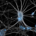
A Cortical Neuron In Conversation.jpg 512 × 512; 38 KB
-
A Cortical Neuron In Isolation.jpg 512 × 512; 55 KB
-
ABC-method.png 557 × 434; 68 KB
-
All Together.jpg 647 × 560; 298 KB
-
ANCA ETHANOL AND FORMALIN.JPEG 2,918 × 2,189; 5.71 MB
-
C anca.jpg 372 × 393; 144 KB
-
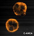
C ANCA.jpg 2,048 × 1,536; 2.77 MB
-
Cell flower-1.jpg 1,376 × 1,032; 134 KB
-
Cell membrane.jpg 1,529 × 1,181; 426 KB
-
Cell vessels.jpg 1,292 × 1,181; 438 KB
-
CONDENSING CHROMOSOMES 2.jpg 1,935 × 549; 785 KB
-
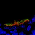
Corneal epithélium.jpg 1,376 × 1,032; 171 KB
-
Cortical Neuron Synapses.jpg 512 × 512; 62 KB
-
Cytokeratin 8.jpg 1,986 × 2,238; 1.89 MB
-
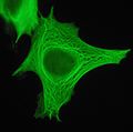
Cytokeratin filaments.jpg 1,692 × 1,676; 1.14 MB
-
DsDNA Abs 2.jpg 718 × 660; 286 KB
-
DsDNA Abs 3.jpg 1,106 × 1,050; 1.2 MB
-
F-actin filaments in cardiomyocytes.jpg 1,600 × 1,200; 269 KB
-
Fish or snail?.jpg 681 × 367; 188 KB
-
Flagella in thymus.jpg 1,024 × 1,024; 123 KB
-
Hartsock Immunological Methods Immunofluorescence.png 350 × 162; 18 KB
-
Heart ece.jpg 720 × 960; 25 KB
-
HEK 293.jpg 4,785 × 4,620; 10.51 MB
-
Hep2IntermediateFilaments2.JPG 687 × 583; 87 KB
-
HIF1a-induziert-Zellkern-rot-durchCOCl2.png 1,300 × 1,030; 863 KB
-
Homologous recombination in intestinal zebrafish tissue.jpg 1,672 × 1,349; 183 KB
-
Honeycomb cells-2.jpg 1,376 × 1,032; 529 KB
-
Honeycomb cells.jpg 1,376 × 1,032; 142 KB
-
HSP IF IgA.jpg 600 × 450; 46 KB
-
Human Cell.jpg 2,801 × 2,446; 3.43 MB
-
IFI-Nuclear and inespecific citoplasmatic stain Hep-2 Line cells.jpg 1,476 × 1,118; 1.1 MB
-
Immunofluorescence light microscopy I.jpg 1,375 × 1,375; 770 KB
-
Immunofluorescence light microscopy II.jpg 1,526 × 1,200; 586 KB
-
Immunofluorescence Mechanism .png 720 × 405; 42 KB
-
Immunofluorescence(Commons).tif 720 × 405; 858 KB
-
Immunofluorescence.jpg 5,532 × 2,907; 1.36 MB
-
Immunofluorescent Stain FAM149B1.jpg 800 × 800; 111 KB
-
Ki-67 protein.jpg 2,118 × 2,388; 2.6 MB
-
LSAB-method.jpg 489 × 375; 35 KB
-
MAP2-tau in neurons.jpg 926 × 935; 1.56 MB
-
Metaphase chromosomes.jpg 399 × 192; 55 KB
-
Microglia and neurons.jpg 1,600 × 1,200; 258 KB
-
Neuron in tissue culture.jpg 1,315 × 1,033; 410 KB
-
Neuronal Dendrites Finding A Path.jpg 512 × 512; 77 KB
-
NUCLEAR DOTS.jpg 1,030 × 840; 1.11 MB
-
Oct4KOT1DAPIT2Acta2T3Oct4T4eYFP1 500IgGOct4IgGeYFP 3.tif 512 × 512; 769 KB
-
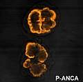
P anca.jpg 347 × 343; 129 KB
-
P ANCA.jpg 2,048 × 1,536; 1.96 MB
-
Parasite150075-fig2 Toxoplasma gondii in Giant panda.tif 1,171 × 883, 2 pages; 281 KB
-
Physical cell communications.jpg 2,362 × 1,772; 919 KB
-
Picture1g.png 297 × 185; 78 KB
-
Primary Immunofluorescence.png 722 × 764; 104 KB
-
Proliferation in situ.jpg 1,031 × 1,031; 53 KB
-
Secondary Immunofluorescence.png 786 × 1,096; 152 KB
-
Speckled HEp-2 cells, immunofluorescence (16099409590).jpg 1,257 × 1,257; 940 KB
-
SPECKLED.jpg 873 × 711; 327 KB
-
Spectrin localization under the neuronal plasme membrane..jpg 800 × 600; 166 KB
-
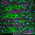
Spinal cord gray matter immunofluorescence staining, confocal imaging.png 1,600 × 1,600; 5.88 MB
-
Swan?.jpg 351 × 705; 230 KB
-
Tela de araña.jpg 857 × 321; 272 KB
-
The Galaxy Within.jpg 1,392 × 1,040; 2.04 MB
-
The Universe Within.jpg 1,058 × 1,211; 164 KB
-
ToxoTachyzoitesGreen.jpg 300 × 300; 3 KB
-
Vimentin and Nuclear Pore Complexes in HeLa cells.jpg 1,600 × 1,200; 183 KB
-
Vimentin.jpg 2,304 × 3,072; 2.06 MB
-
Αστροκύτταρα ποντικού σε καλλιέργεια.jpg 1,024 × 1,024; 120 KB
-
Ιππόκαμπος ποντικού.jpg 1,024 × 1,024; 148 KB
-
Πολλαπλασιασμός νευρικών βλαστικών κυττάρων ποντικών.jpg 924 × 836; 113 KB
