Category:Images translated by Alaa
Jump to navigation
Jump to search
Media in category "Images translated by Alaa"
The following 200 files are in this category, out of 283 total.
(previous page) (next page)-
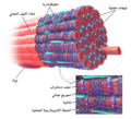
1023 T-tubule-ar.jpg 577 × 368; 83 KB
-
1610 Muscles Controlled by the Accessory Nerve-02-ar.jpg 1,299 × 1,081; 264 KB
-
20090428MSLEntry1-ar.jpg 640 × 390; 85 KB
-
2313 The Lung Pleurea-ar.jpg 1,925 × 1,450; 433 KB
-
2424 Exocrine and Endocrine Pancreas-ar.jpg 637 × 827; 238 KB
-
2425 Gallbladder-ar.jpg 787 × 641; 199 KB
-
2604 Nerves Innervating the Urinary SystemN-ar.jpg 1,738 × 1,265; 761 KB
-
2917 Size of Uterus Throughout Pregnancy-02-ar.jpg 449 × 599; 56 KB
-
2919 Hormones Initiating Labor-02-ar.jpg 800 × 449; 52 KB
-
3D Medical Animation Bronchial Airways terminating ends-ar.jpg 1,920 × 1,080; 436 KB
-
3D medical animation corona virus-ar.jpg 1,920 × 1,080; 458 KB
-
3D medical animation coronavirus structure-ar.jpg 1,920 × 1,080; 489 KB
-
404 Goblet Cell new-ar.jpg 2,539 × 4,722; 2.13 MB
-
A Course of Shingles diagram-ar.png 488 × 477; 113 KB
-
Abdomen-periumbilical region-ar.png 545 × 574; 227 KB
-
Abscess diag 02-ar.svg 320 × 199; 315 KB
-
Alveolus diagram-ar.png 418 × 312; 63 KB
-
Amyloid-plaque formation-big-ar.jpg 2,844 × 630; 758 KB
-
Anatomy Abdomen Tiesworks-ar.jpg 642 × 599; 72 KB
-
Anatomy and physiology of animals A reflex arc-ar.jpg 549 × 272; 96 KB
-
Anthropornis v1-ar.svg 484 × 289; 201 KB
-
Asteroid2000EM26-NearEarthEncounter-20140217-ar.png 581 × 304; 86 KB
-
Asthma attack-illustration NIH-ar.jpg 475 × 315; 111 KB
-
Axial skeleton diagram-ar.svg 427 × 562; 938 KB
-
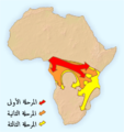
Bantu expansion-ar.png 400 × 424; 138 KB
-
Base of skull 19-ar.jpg 960 × 720; 350 KB
-
Biliary system new-ar.svg 631 × 587; 418 KB
-
Bipolar mood shifts-ar.png 955 × 1,191; 419 KB
-
BJS Sexual Assualt Rates 1995-2013-ar.png 508 × 435; 29 KB
-
Blausen 0004 AbdominalParacentesis-ar.png 600 × 600; 374 KB
-
Blausen 0216 CerebrospinalSystem-ar.png 800 × 600; 503 KB
-
Blausen 0257 CoronaryArtery Plaque-ar.png 940 × 661; 772 KB
-
Blausen 0399 FemaleReproSystem 01-ar.png 600 × 600; 192 KB
-
Blausen 0400 FemaleReproSystem 02-ar.png 733 × 600; 589 KB
-
Blausen 0406 FingerNailAnatomy-ar.png 750 × 1,500; 717 KB
-
Blausen 0463 HeartAttack-ar.png 450 × 600; 220 KB
-
Blausen 0470 HeartWall-ar.png 600 × 600; 477 KB
-
Blausen 0699 PancreasAnatomy2-ar.png 750 × 600; 386 KB
-
Blausen 0701 PancreaticTissue-ar.png 600 × 600; 372 KB
-
Blausen 0742 Pneumothorax-ar.png 1,500 × 1,500; 1.7 MB
-
Blausen 0785 Scoliosis 01-ar.png 300 × 600; 296 KB
-
Blausen 0801 SkeletalMuscle-ar.png 2,000 × 1,823; 4.84 MB
-
Blausen 0805 Skin MerkelCell-ar.png 800 × 640; 513 KB
-
Brain human normal inferior view with labels ar.svg 424 × 505; 244 KB
-
Brain size comparison - Brain size (gram)-ar.png 670 × 473; 21 KB
-
Brantigan 1963 1-53-ar.png 735 × 1,503; 98 KB
-
BristolStoolChart-ar.png 1,196 × 720; 302 KB
-
Buccal Fat Diagram-ar.jpg 847 × 759; 66 KB
-
Bumm 123 lg-ar.jpg 800 × 598; 126 KB
-
Bumm 158 lg-ar.jpg 473 × 599; 103 KB
-
Carboxysome-ar.png 3,558 × 1,356; 5.79 MB
-
Carpal Tunnel Syndrome-ar.png 1,500 × 1,500; 2.19 MB
-
Carte Coffea robusta arabic-ar.png 4,500 × 2,223; 1.06 MB
-
Cellular Fluid Content-ar.jpg 399 × 385; 29 KB
-
Ch4 kinases-ar.jpg 380 × 279; 28 KB
-
Change in Average Temperature-ar.svg 960 × 816; 709 KB
-
Children's pain scale-ar.jpg 1,042 × 404; 57 KB
-
Chylomicronarabic.jpg 500 × 262; 85 KB
-
Clitoris inner anatomy-ar.png 630 × 650; 87 KB
-
Clitoris outer anatomy-ar.png 585 × 619; 68 KB
-
Clonorchis sinensis LifeCycle-ar.png 562 × 435; 54 KB
-
Colonizationoftheamericas-ar.png 463 × 599; 68 KB
-
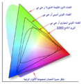
Colorspace-ar.png 661 × 679; 456 KB
-
Cornea-ar.png 452 × 464; 58 KB
-
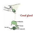
Coxal-gland.jpg 1,000 × 1,000; 140 KB
-
Culex pipiens diagram ar.svg 512 × 539; 684 KB
-
Decompressive Craniectomy-ar.png 1,829 × 1,856; 951 KB
-
Dengue testing-ar.png 829 × 565; 28 KB
-
Depiction of a woman suffering from Osteopenia-ar.png 2,605 × 1,667; 911 KB
-
Diagram human cell nucleus ar.svg 462 × 378; 313 KB
-
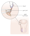
Diagram showing a cystoscopy for a man and a woman CRUK 064-ar.png 375 × 555; 72 KB
-
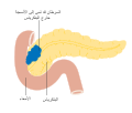
Diagram showing stage T3 cancer of the pancreas CRUK 261-ar.svg 375 × 320; 90 KB
-
Diagram showing stage T4 cancer of the pancreas CRUK 267-ar.svg 375 × 321; 117 KB
-
Diagram showing the position of the pancreas CRUK 356-ar.svg 375 × 339; 241 KB
-
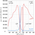
Diel-ar.png 361 × 277; 14 KB
-
Digestive system showing bile duct-ar.png 300 × 222; 9 KB
-
Dorsiplantar-ar.jpg 611 × 874; 85 KB
-
Duodenumandpancreas-ar.jpg 1,050 × 766; 168 KB
-
Fatimid Caliphate-ar.png 800 × 459; 61 KB
-
Female and Male Urethra-ar.jpg 454 × 600; 147 KB
-
Figure 28 01 06-ar.jpg 781 × 718; 208 KB
-
Figure 28 02 01-ar.JPG 555 × 789; 181 KB
-
Figure 28 02 06-ar.jpg 818 × 393; 171 KB
-
Fingernail label-ar.jpg 640 × 480; 62 KB
-
Flowchart of Stewards election-ar.svg 638 × 709; 210 KB
-
Formation of Cellulite-ar.jpg 720 × 539; 125 KB
-
Gallbladder (organ)-ar.png 1,200 × 1,200; 1.18 MB
-
Global Temperature And Forces-ar.svg 1,400 × 1,050; 601 KB
-
Glycogen phosphorylase2-ar.png 799 × 173; 30 KB
-
Goblin shark size-ar.svg 3,115 × 1,435; 58 KB
-
Grant 1962 214-ar.png 2,651 × 3,168; 1.38 MB
-
Grant 1962 215-ar.png 2,519 × 1,969; 1.29 MB
-
Gray 1100 Pancreatic duct-ar.png 600 × 427; 109 KB
-
Gray100-ar.png 230 × 599; 34 KB
-
Gray1034-ar.png 461 × 800; 188 KB
-
Gray1097-ar.png 800 × 595; 327 KB
-
Gray1100-ar.png 600 × 427; 107 KB
-
Gray1101-ar.png 400 × 322; 25 KB
-
Gray1102-ar.png 400 × 305; 27 KB
-
Gray1105-ar.png 426 × 400; 167 KB
-
Gray1113-ar.png 600 × 373; 77 KB
-
Gray112-ar.png 436 × 600; 126 KB
-
Gray1138-ar.png 517 × 350; 57 KB
-
Gray1146-ar.png 452 × 400; 102 KB
-
Gray1152-ar.png 398 × 400; 106 KB
-
Gray1154-ar.png 389 × 700; 116 KB
-
Gray1156-ar.png 730 × 700; 320 KB
-
Gray1160-ar.png 437 × 400; 69 KB
-
Gray1161-ar.png 527 × 350; 116 KB
-
Gray1164-ar.png 400 × 350; 59 KB
-
Gray1165-ar.png 671 × 500; 185 KB
-
Gray1166-ar.png 600 × 600; 192 KB
-
Gray1167-ar.png 708 × 768; 115 KB
-
Gray1169-ar.png 597 × 450; 117 KB
-
Gray1170-ar.png 660 × 500; 119 KB
-
Gray1204-ar-v2.png 511 × 400; 86 KB
-
Gray1204-ar.png 511 × 400; 84 KB
-
Gray1205-ar.png 429 × 350; 70 KB
-
Gray1220-ar.svg 434 × 550; 150 KB
-
Gray1225-ar.png 508 × 400; 82 KB
-
Gray1229-ar.png 429 × 500; 123 KB
-
Gray1230-ar.png 581 × 500; 132 KB
-
Gray1238-ar.png 311 × 500; 104 KB
-
Gray242-ar.png 450 × 321; 72 KB
-
Gray303-ar.png 349 × 350; 55 KB
-
Gray314-ar.png 270 × 553; 28 KB
-
Gray332-ar.png 249 × 550; 84 KB
-
Gray34-ar.png 500 × 383; 66 KB
-
Gray408-ar.png 460 × 500; 133 KB
-
Gray413 color-ar.png 550 × 396; 61 KB
-
Gray420-ar.png 329 × 550; 81 KB
-
Gray432-ar.png 683 × 700; 199 KB
-
Gray488-ar.png 450 × 359; 53 KB
-
Gray535-ar.png 395 × 500; 99 KB
-
Gray543-ar.png 700 × 569; 186 KB
-
Gray588-ar.png 359 × 277; 39 KB
-
Gray589-ar.png 600 × 356; 111 KB
-
Gray614-ar.png 450 × 523; 115 KB
-
Gray662-ar.png 260 × 600; 60 KB
-
Gray791-ar.png 449 × 450; 51 KB
-
Gray794-ar.png 638 × 700; 258 KB
-
Gray803-ar.png 576 × 500; 205 KB
-
Gray82-ar.png 450 × 287; 38 KB
-
Gray828-ar.png 493 × 650; 69 KB
-
Gray849-ar.png 600 × 664; 193 KB
-
Gray881-ar.png 500 × 335; 52 KB
-
Gray90-ar.png 450 × 344; 42 KB
-
Gray944-ar.png 600 × 511; 131 KB
-
Gray95-ar.png 525 × 550; 157 KB
-
Gray954-ar-v2.png 373 × 600; 123 KB
-
Gray954-ar.png 373 × 600; 121 KB
-
Gray956-ar.png 450 × 354; 53 KB
-
Gray96-ar.png 567 × 500; 105 KB
-
Gray967-ar.png 600 × 434; 144 KB
-
Gray97-ar.png 381 × 600; 89 KB
-
Gray98-ar.png 500 × 333; 92 KB
-
HarmCausedByDrugsTable-ar.png 794 × 794; 44 KB
-
Heartfailure-ar.jpg 377 × 500; 42 KB
-
Hematopoesis Ar.svg 798 × 382; 2.15 MB
-
Herd immunity-ar.png 2,000 × 2,456; 1.08 MB
-
Herpes zoster ophthalmicus.2-ar.jpg 752 × 720; 176 KB
-
How big is 1 micrometer (10690468113)-ar.jpg 5,506 × 6,886; 4.71 MB
-
Human photoreceptor distribution-ar.png 2,048 × 2,048; 265 KB
-
Ileal pouch-anal anastomosis-ar.svg 460 × 328; 180 KB
-
Illu bladder-ar.jpg 520 × 273; 57 KB
-
Illu blood components-ar.png 506 × 416; 14 KB
-
Illu breast anatomy-ar.jpg 274 × 349; 49 KB
-
Illu cervix-ar.jpg 496 × 293; 61 KB
-
Illu ovaryb-ar.jpg 319 × 168; 24 KB
-
Illu penis-ar.jpg 520 × 300; 87 KB
-
Illu prostate lobes-ar.jpg 400 × 250; 37 KB
-
Illu prostate zones-ar.jpg 367 × 226; 35 KB
-
Illu pulmonary circuit-ar.jpg 520 × 250; 46 KB
-
Illu repdt female-ar.jpg 520 × 300; 77 KB
-
Illu skin02-ar.jpg 397 × 302; 39 KB
-
Illu upper extremity-ar.jpg 350 × 231; 39 KB
-
Illu urinary system-ar.jpg 356 × 304; 27 KB
-
Illu vertebral column-ar.jpg 350 × 350; 43 KB
-
Illu04 tongue-ar.jpg 500 × 326; 79 KB
-
In situ carcinoma-ar.jpg 380 × 283; 27 KB
-
Intracranial electrode grid for electrocorticography-ar.png 1,500 × 1,500; 1.11 MB
-
Larynx external-ar.svg 1,125 × 975; 303 KB
-
Lawrence 1960 17.26-ar.png 2,052 × 1,276; 455 KB
-
Leukemia- SAG-ar.jpg 1,920 × 1,080; 322 KB
-
Life Cycle of the Malaria Parasite-ar.jpg 1,300 × 1,282; 730 KB
-
Ligature-ar.jpg 1,629 × 1,122; 179 KB
-
Limbus-ar.png 452 × 464; 60 KB
-
Locus Kiesselbachii Shematic-ar.jpg 634 × 465; 134 KB
-
Lungs open-ar.jpg 350 × 292; 58 KB
-
Lymphatics of the prostate-Gray619-ar.png 701 × 648; 201 KB
-
Mashreq-ar.png 379 × 376; 14 KB
-
Menkes disease3-ar.jpg 600 × 428; 111 KB
-
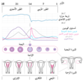
MenstrualCycle2-ar.png 430 × 420; 47 KB


