Category:Human temporal bones
Jump to navigation
Jump to search
Subcategories
This category has the following 30 subcategories, out of 30 total.
3
A
- Articular tubercle (18 F)
H
I
- Internal acoustic meatus (8 F)
J
L
M
- Mandibular fossa (33 F)
- Mastoid cells (10 F)
- Mastoid notch (14 F)
P
S
- Sheath of styloid process (20 F)
- Sigmoid sulcus (12 F)
- Sphenosquamosal sutures (26 F)
- Squamosal sutures (41 F)
T
V
Z
- Zygomatic arch (21 F)
- Zygomaticotemporal sutures (27 F)
Media in category "Human temporal bones"
The following 171 files are in this category, out of 171 total.
-

-
 708 Temporal Bone.jpg 774 × 555; 177 KB
-
A-Surgical-Navigation-System-for-Guiding-Exact-Cochleostomy-Using-Auditory-Feedback-A-Clinical-769659.f1.ogv 43 s, 1,440 × 1,080; 5.6 MB
-
 Base of skull 17.jpg 960 × 720; 108 KB
-
 BodyParts3D FJ6478 FJ6391 Temporal bone.stl 5,120 × 2,880; 479 KB
-
 Braus 1921 328.png 1,392 × 1,144; 4.56 MB
-
 Braus 1921 340.png 1,464 × 996; 4.18 MB
-
 Braus 1921 341.png 1,512 × 1,152; 4.99 MB
-
 Braus 1921 342.png 1,520 × 884; 3.85 MB
-
 Braus 1921 343.png 1,536 × 1,092; 4.81 MB
-
 Braus 1921 344.png 1,432 × 1,104; 4.53 MB
-
 Braus 1921 345.png 1,344 × 976; 3.76 MB
-
 Braus 1921 346.png 512 × 740; 1.09 MB
-
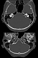
 Canalis petromastoideus 58M - CT axial - 001 - Annotation.jpg 683 × 850; 61 KB
-
 Canalis petromastoideus 58M - CT axial - 001.jpg 683 × 850; 60 KB
-
 Canalis petromastoideus 78M - CT axial - 001 - Annotation.jpg 1,092 × 1,109; 98 KB
-
 Canalis petromastoideus 78M - CT axial - 001.jpg 1,092 × 1,109; 97 KB
-
 Canalis petromastoideus 85M - CT axial - 001 - Annotation.jpg 881 × 1,082; 83 KB
-
 Canalis petromastoideus 85M - CT axial - 001.jpg 881 × 1,082; 82 KB
-
 Canalis subarcuatis 86W - CT axial - 001.jpg 1,171 × 1,394; 147 KB
-
 ConduitOsseux.jpg 2,061 × 1,532; 1.44 MB
-
 Cranio. Norma laterale. Splancnocranio.jpg 960 × 720; 84 KB
-
 Cranium - lateral view.jpg 4,608 × 3,456; 4.79 MB
-
 Cranium - os temporale (intra lateralis) 2.jpg 4,608 × 3,456; 4.19 MB
-
 Cranium - os temporale (intra lateralis) 3.jpg 4,608 × 3,456; 5.1 MB
-
 Cranium - os temporale (intra lateralis).jpg 3,456 × 4,608; 4.43 MB
-
 Cranium - os temporale (lateralis).jpg 3,456 × 4,608; 4.83 MB
-
 Cranium - os temporale (lateralis, dexter) 2.jpg 4,608 × 3,456; 4.53 MB
-
 Cranium - os temporale (lateralis, dexter).jpg 3,456 × 4,608; 4.29 MB
-
 Cranium - os temporale (meatus acusticus externus detail, lateral).jpg 3,456 × 4,608; 3.5 MB
-
 Cunningham’s Text-book of Anatomy (1914) - Fig 143.png 2,144 × 802; 1.39 MB
-
 Dixon's Manual of human osteology (1912) - Fig 104.png 1,478 × 1,088; 676 KB
-
 File Crane déformé 1905 MHNT (droite).jpg 3,888 × 2,954; 5.92 MB
-
 Gerrish's Text-book of Anatomy (1902) - Fig. 211.png 1,314 × 1,072; 953 KB
-
 Gerrish's Text-book of Anatomy (1902) - Fig. 212.png 1,334 × 1,188; 1.03 MB
-
 Gerrish's Text-book of Anatomy (1902) - Fig. 213.png 726 × 1,152; 763 KB
-
 Gerrish's Text-book of Anatomy (1902) - Fig. 214.png 1,270 × 1,344; 995 KB
-
 Gerrish's Text-book of Anatomy (1902) - Fig. 215.png 564 × 604; 203 KB
-
 Gray 137 - Os temporal gauche - vue externe.png 500 × 449; 208 KB
-
 Gray 138 - Os temporal gauche - Vue médiale.png 573 × 550; 272 KB
-
 Gray 141 - Os temporal gauche - Vue inférieure.png 550 × 495; 289 KB
-
 Gray137.png 500 × 449; 41 KB
-
 Gray137az.png 502 × 449; 106 KB
-
 Gray138.png 573 × 550; 66 KB
-
 Gray139.png 550 × 502; 53 KB
-
 Gray140.png 319 × 304; 27 KB
-
 Gray141.png 550 × 495; 34 KB
-
 Gray142.png 500 × 327; 20 KB
-
 Gray143.png 431 × 357; 20 KB
-
 Gray143az.png 431 × 357; 79 KB
-
 Gray144.png 329 × 364; 20 KB
-
 Gray144az.png 329 × 364; 81 KB
-
 Gray164-ar.png 400 × 379; 191 KB
-
 Gray164.png 400 × 379; 49 KB
-
 Gray187.png 718 × 1,169; 147 KB
-
 Gray188 no text color temporal r.jpg 716 × 537; 120 KB
-
 Gray193.png 719 × 1,057; 150 KB
-
 Gray194.png 650 × 420; 84 KB
-
 Gray194az.png 650 × 420; 284 KB
-
 Gray309-ar.svg 471 × 456; 401 KB
-
 Gray309.png 450 × 435; 39 KB
-
 Gray913.png 550 × 492; 51 KB
-
 Gray922.png 500 × 463; 56 KB
-
 Gray923.png 600 × 434; 61 KB
-
 Gray995 zh.png 2,504 × 2,036; 2.31 MB
-
 Gray995.png 2,504 × 2,036; 2.82 MB
-
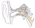
 Hearing mechanics.jpg 614 × 650; 249 KB
-
 Holden's human osteology (1899) - Plt08 Fig01.png 1,912 × 1,420; 2.29 MB
-
 Holden's human osteology (1899) - Plt08 Fig02.png 1,881 × 1,299; 1.8 MB
-
 Holden's human osteology (1899) - Plt09 Fig01.png 1,584 × 1,352; 1.04 MB
-
 Holden's human osteology (1899) - Plt09 Fig02.png 1,792 × 1,128; 1.79 MB
-
 Holden's human osteology (1899) - Plt21.png 1,678 × 2,805; 2.28 MB
-
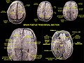 Horizontal sections of fetal brain.jpg 960 × 720; 123 KB
-
 Huesotemporal.svg 1,052 × 531; 919 KB
-
 Human skull 2008.jpg 1,936 × 1,288; 1.71 MB
-
 Left temporal bone close-up anterior.png 900 × 900; 89 KB
-
 Left temporal bone close-up inferior animation.gif 320 × 320; 752 KB
-
 Left temporal bone close-up inferior.png 900 × 900; 141 KB
-
 Left temporal bone close-up lateral animation.gif 320 × 320; 684 KB
-
 Left temporal bone close-up lateral animation2.gif 450 × 450; 1.03 MB
-
 Left temporal bone close-up lateral.png 900 × 900; 116 KB
-
 Left temporal bone close-up medial.png 900 × 900; 153 KB
-
 Left temporal bone close-up posterior.png 900 × 900; 99 KB
-
 Left temporal bone close-up superior animation.gif 320 × 320; 666 KB
-
 Left temporal bone close-up superior.png 900 × 900; 117 KB
-
 Mandibular fossa 3.jpg 960 × 720; 113 KB
-
 Mastoid process.jpg 960 × 720; 111 KB
-
 Morris' human anatomy (1898) - Fig 047.png 1,164 × 834; 636 KB
-
 Morris' human anatomy (1898) - Fig 049.png 1,623 × 576; 689 KB
-
 Morris' human anatomy (1933) - Fig 164.png 2,169 × 1,326; 2.68 MB
-
 Morris' human anatomy (1933) - Fig 165.png 1,954 × 1,396; 1.76 MB
-
 Morris' human anatomy (1933) - Fig 167.png 1,965 × 1,647; 1.84 MB
-
 Morris' human anatomy (1933) - Fig 168.png 2,028 × 688; 1.14 MB
-
 Occipitomastoid suture.png 1,600 × 1,200; 52 KB
-
 OreilleMoyenneSchema.jpg 3,697 × 2,901; 2.2 MB
-
 Petrosquamózusvarrat.PNG 329 × 364; 33 KB
-
 Piersol's human anatomy (1919) - Fig. 195.png 1,494 × 1,128; 1.07 MB
-
 Piersol's human anatomy (1919) - Fig. 196.png 1,316 × 1,230; 558 KB
-
 Piersol's human anatomy (1919) - Fig. 197.png 1,434 × 1,040; 1.02 MB
-
 Piersol's human anatomy (1919) - Fig. 198.png 1,258 × 1,056; 564 KB
-
 Piersol's human anatomy (1919) - Fig. 199.png 1,512 × 1,030; 660 KB
-
 Piersol's human anatomy (1919) - Fig. 200.png 1,491 × 1,006; 740 KB
-
 Piersol's human anatomy (1919) - Fig. 201.png 829 × 567; 213 KB
-
 Piersol's human anatomy (1919) - Fig. 202.png 788 × 789; 115 KB
-
 Piersol's human anatomy (1919) - Fig. 203.png 1,275 × 1,071; 662 KB
-
 Piersol's human anatomy (1919) - Fig. 204.png 1,035 × 817; 271 KB
-
 Piersol's human anatomy (1919) - Fig. 205.png 796 × 632; 243 KB
-
 Piersol's human anatomy (1919) - Fig. 206.png 942 × 588; 268 KB
-
 Piersol's human anatomy (1919) - Fig. 207.png 736 × 452; 147 KB
-
 Pneumatisierte Felsenbeinspitze 86jw - CT axial - 001.jpg 1,086 × 1,647; 157 KB
-
 Processus styloideus (close).png 636 × 458; 483 KB
-
 Processus styloideus.jpg 1,122 × 1,095; 134 KB
-
 Pterygopalatine fossa.PNG 640 × 480; 743 KB
-
 Rocher global.jpg 13,000 × 6,500; 13.82 MB
-
 Rotation temporal bone.gif 600 × 600; 3.25 MB
-
 SchaedelSeitlichSutur10.png 1,600 × 1,200; 32 KB
-
 SchaedelSeitlichSutur3.png 1,600 × 1,200; 33 KB
-
 SchaedelSeitlichSutur4.png 1,600 × 1,200; 32 KB
-
 Slide8STE.JPG 960 × 720; 72 KB
-
 Sobo 1909 55.png 2,172 × 1,420; 8.84 MB
-
 Sobo 1909 56.png 2,132 × 1,484; 9.07 MB
-
 Sobo 1909 57.png 2,196 × 1,360; 8.56 MB
-
 Sobo 1909 58.png 1,572 × 1,488; 6.7 MB
-
 Sobo 1909 59.png 1,072 × 880; 2.7 MB
-
 Sobo 1909 60.png 1,232 × 956; 3.38 MB
-
 Sobo 1909 61.png 1,856 × 1,032; 5.49 MB
-
 Sobo 1909 62.png 1,960 × 1,076; 6.04 MB
-
 Sobo 1909 63.png 2,112 × 1,016; 6.15 MB
-
 Sobo 1909 781.png 2,272 × 2,216; 1.99 MB
-
 Sotokawazu03.jpg 550 × 378; 43 KB
-
 Sphenoid and temporal bones 2.jpg 960 × 720; 88 KB
-
 Sphenoid and temporal bones 3.jpg 960 × 720; 88 KB
-
 Sphenoid and temporal bones 4.jpg 960 × 720; 94 KB
-
 Sphenoid and temporal bones.jpg 960 × 720; 91 KB
-
 Temporal (10819026425).jpg 1,872 × 2,689; 708 KB
-
 Temporal (10825825186).jpg 2,057 × 2,025; 794 KB
-
 Temporal bone - animation 01.gif 600 × 600; 20.25 MB
-
 Temporal bone - animation 02.gif 600 × 600; 21.62 MB
-
 Temporal bone - The Hungry Artist Multimedia.jpg 2,500 × 2,500; 1.54 MB
-
 Temporal bone anterior.png 900 × 900; 178 KB
-
 Temporal bone anterior2.png 900 × 900; 131 KB
-
 Temporal bone inferior animation.gif 320 × 320; 971 KB
-
 Temporal bone inferior.png 900 × 900; 216 KB
-
 Temporal bone inferior2.png 900 × 900; 218 KB
-
 Temporal bone lateral animation.gif 320 × 320; 929 KB
-
 Temporal bone lateral.png 900 × 900; 150 KB
-
 Temporal bone lateral2.png 900 × 900; 164 KB
-
 Temporal bone lateral3.png 900 × 900; 157 KB
-
 Temporal bone lateral4.png 900 × 900; 210 KB
-
 Temporal bone lateral5.png 4,500 × 4,500; 3.93 MB
-
 Temporal bone posterior.png 900 × 900; 135 KB
-
 Temporal Bone Simple.png 600 × 600; 61 KB
-
 Temporal bone superior animation.gif 320 × 320; 692 KB
-
 Temporal bone superior animation2.gif 320 × 320; 833 KB
-
 Temporal bone superior.png 900 × 900; 211 KB
-
 Temporal bone.png 800 × 455; 298 KB
-
 Temporal bone2.jpg 2,406 × 1,805; 2.03 MB
-
 Temporal bone3.jpg 3,168 × 2,376; 3.32 MB
-
 Temporal droit.png 727 × 534; 150 KB
-
 Temporal fossa.jpg 960 × 720; 87 KB
-
 Temporo-parietal suture 3.jpg 960 × 720; 93 KB
-
 Temporo-parietal suture 4.jpg 960 × 720; 73 KB
-
 Temporo-parietal suture.jpg 960 × 720; 85 KB
-
 Temporomandibular joint.jpg 960 × 720; 63 KB
-
 Testut's Treatise on Human Anatomy (1911) - Vol 1 - Fig 166.png 1,512 × 1,434; 1.8 MB
-
 Tuba auditiva in der Computertomografie 72M - CT axial - 001 - Annotation.jpg 1,436 × 1,109; 101 KB
-
 Tuba auditiva in der Computertomografie 72M - CT axial - 001.jpg 1,436 × 1,109; 91 KB
-
Validation-of-Exposure-Visualization-and-Audible-Distance-Emission-for-Navigated-Temporal-Bone-pone.0041262.s001.ogv 1 min 46 s, 640 × 480; 17.73 MB
-
 Vue antérieure de l'os temporal.png 1,920 × 1,920; 1.39 MB
-
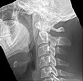
 X-ray manual - U.S. Army (1917) (14570635290).jpg 2,416 × 1,984; 416 KB
-
 Zygomatic arch.jpg 960 × 720; 115 KB


_2.jpg)
_3.jpg)
.jpg)
.jpg)
_2.jpg)
.jpg)
.jpg)
_-_Fig_104.png)
.jpg)
_-_Fig._211.png)
_-_Fig._212.png)
_-_Fig._213.png)
_-_Fig._214.png)
_-_Fig._215.png)


_-_Plt08_Fig01.png)
_-_Plt08_Fig02.png)
_-_Plt09_Fig01.png)
_-_Plt09_Fig02.png)
_-_Plt21.png)


_-_Fig_047.png)
_-_Fig_164.png)
_-_Fig_165.png)
_-_Fig_167.png)


_-_Fig._195.png)
_-_Fig._196.png)
_-_Fig._197.png)
_-_Fig._198.png)
_-_Fig._199.png)
_-_Fig._200.png)
_-_Fig._201.png)
_-_Fig._202.png)
_-_Fig._203.png)
_-_Fig._204.png)
_-_Fig._205.png)
_-_Fig._206.png)
_-_Fig._207.png)

.png)


.jpg)
.jpg)


_-_Vol_1_-_Fig_166.png)


_(14570635290).jpg)

_-_Fig_143.png)
_-_Fig_049.png)
_-_Fig_168.png)