Category:Human teeth
Jump to navigation
Jump to search
calcified whitish structure in humans' mouths used to break down food | |||||
| Upload media | |||||
| Instance of |
| ||||
|---|---|---|---|---|---|
| Subclass of | |||||
| Part of | |||||
| Has part |
| ||||
| |||||
Subcategories
This category has the following 36 subcategories, out of 36 total.
*
B
- BL02-J54-100 (7 F)
C
D
- Dental nomenclature (18 F)
F
G
H
I
- Human incisors (34 F)
M
- Mamelon (dentistry) (1 F)
P
S
- Sharpened teeth (33 F)
T
- Teeth needing sanitation (14 F)
- Teeth resorption (9 F)
W
Y
- Yaeba (4 F)
Media in category "Human teeth"
The following 113 files are in this category, out of 113 total.
-
2017-04-29-IMG 6888-Skull (Cut out).jpg 2,448 × 3,264; 2 MB
-
3D Medical Animation Still Showing Types of Teeth.jpg 1,426 × 1,080; 521 KB
-
3D model of the human mouth.stl 5,120 × 2,880; 2.18 MB
-
3D partial human mandible with teeth.stl 5,120 × 2,880; 14.74 MB
-
Alveolar processes cutaway color.jpg 1,480 × 1,271; 1.5 MB
-
Arbetsliv.jpg 3,799 × 2,618; 4.99 MB
-
ARQUITECTURA POSITIVA.png 586 × 541; 600 KB
-
ARQÓSEA.png 1,213 × 318; 292 KB
-
AT1.png 1,929 × 1,419; 1.44 MB
-
Baby tooth 1 (5614978435).jpg 2,048 × 2,240; 361 KB
-
Balance-schiene-am-patienten-1.jpg 1,500 × 1,000; 469 KB
-
Balance-schiene-am-patienten-2.jpg 1,500 × 1,000; 385 KB
-
Bleecker st. (2987739690).jpg 1,000 × 662; 117 KB
-
Bleibende Zähne beim Menschen eingefärbt.png 284 × 500; 103 KB
-
BodyParts3D Tooth.stl 5,120 × 2,880; 1.9 MB
-
Bondingbefore-after2.jpg 240 × 240; 12 KB
-
Brian diastema.png 1,375 × 993; 1.82 MB
-
Buchenwald Teeth 74565.jpg 470 × 370; 81 KB
-
Canine guidance.jpg 631 × 263; 14 KB
-
Captura de pantalla 2017-11-26 a la(s) 14.29.39.png 297 × 198; 105 KB
-
Captura de pantalla 2017-11-26 a la(s) 18.59.52.png 295 × 191; 118 KB
-
Ceramic braces @15000₹.jpg 209 × 334; 17 KB
-
Ceramics . . . always evocative and inspiring !! (14425627083).jpg 3,264 × 2,448; 2.59 MB
-
Clasificación de furcas.jpg 639 × 381; 96 KB
-
CONFIGURACIÓNÓSEA.jpg 270 × 616; 34 KB
-
Conservative dentistry encias.png 775 × 379; 666 KB
-
Crown core.jpg 2,273 × 1,420; 928 KB
-
Cusp tips.JPG 2,189 × 1,883; 839 KB
-
Cutaway image of human jaw with teeth (1917).jpg 529 × 493; 136 KB
-
Dental problem in 10-year-old girl - 1.jpg 3,120 × 4,160; 3.49 MB
-
Dental problem in 10-year-old girl - 2.jpg 3,120 × 4,160; 2.42 MB
-
Dentition (secondary).png 630 × 579; 71 KB
-
DentRailliet1895MeyCh.jpg 954 × 344; 66 KB
-
Die Gartenlaube (1879) b 507 2.jpg 868 × 875; 142 KB
-
Dientes incisivos en forma de pala.png 536 × 927; 689 KB
-
Distancia y festoneado.png 973 × 557; 1.03 MB
-
DSC 3391 - Flickr - Stiller Beobachter.jpg 2,384 × 2,385; 1.99 MB
-
EXOSTOSISMAXILARSUP.jpg 850 × 418; 69 KB
-
Feldspathic VM9 Porcelain Crowns.jpg 1,280 × 1,280; 111 KB
-
FENESTRACIONYDEHISCENCIA.png 1,020 × 748; 1.34 MB
-
FESTONEADO.png 386 × 542; 427 KB
-
Free Macro White Teeth With Dental Floss and Red Lipstick Creative Commons (509495525).jpg 1,578 × 1,383; 1.25 MB
-
FURCAGRADOIII.png 960 × 634; 1.07 MB
-
FÉRULA DE ADELANTAMIENTO MANDIBULAT.jpg 2,771 × 1,896; 699 KB
-
FÉRULA DE DESCARGA (1).jpg 1,454 × 831; 649 KB
-
FÉRULA DE DESCARGA.jpg 2,840 × 1,931; 869 KB
-
FÉRULA DE REPOSICIONAMIENTO MANDIBULAR.jpg 1,320 × 802; 504 KB
-
Gray187.png 718 × 1,169; 147 KB
-
Gray997 zh-hant.png 284 × 500; 110 KB
-
Gray997 zh.png 284 × 500; 110 KB
-
Gray997-es-dientes.png 284 × 500; 34 KB
-
Gray997.heb.PNG 284 × 500; 34 KB
-
Gray997.png 284 × 500; 34 KB
-
Gray997sv.png 284 × 500; 41 KB
-
GUIA CANINA (FÉRULA DE MICHIGAN).jpg 1,341 × 741; 487 KB
-
Hemifacial microsomia Pruzansky type III.jpg 2,883 × 2,506; 853 KB
-
HK old lady damaged teeth May 2023 01.jpg 1,152 × 2,048; 182 KB
-
HK old lady damaged teeth May 2023 02.jpg 1,152 × 2,048; 164 KB
-
Homo luzonensis Zähne und Fußknochen.jpg 3,173 × 2,617; 1.56 MB
-
Homo sapiens teeth.webp 313 × 322; 11 KB
-
HUESOBULBOSO.png 1,020 × 744; 1.89 MB
-
INVERSAA.png 584 × 384; 545 KB
-
Jointtypodonts.jpg 404 × 251; 38 KB
-
Lips - Flickr - Stiller Beobachter.jpg 2,719 × 2,265; 7.04 MB
-
MateriaAlba.jpg 216 × 149; 4 KB
-
MayanDentalmodification.jpg 918 × 709; 139 KB
-
Molar.png 159 × 150; 12 KB
-
OSTECTOMÍA.png 617 × 220; 77 KB
-
OSTEOPLASTÍA.png 563 × 189; 74 KB
-
Overjet.jpg 1,200 × 688; 91 KB
-
Pieces Of Me (698521).jpeg 2,048 × 1,365; 316 KB
-
PLANA.png 1,298 × 567; 1.12 MB
-
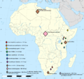
Post-canine Megadontia map in Africa.png 1,525 × 1,440; 566 KB
-
Posterior disocclusion.jpg 538 × 303; 55 KB
-
Premolars.png 159 × 150; 12 KB
-
Read this label your baby's teeth depend on it (6946533407).jpg 1,947 × 3,110; 622 KB
-
Rebekah Radice.jpg 3,840 × 5,760; 14.53 MB
-
Sealants and fluoride (6946537725).jpg 2,238 × 2,717; 651 KB
-
Shedding of first molar teeth at rhe age of 10 years.jpg 4,128 × 3,096; 1.88 MB
-
Solutrense de la Cueva del Parpalló 02.jpg 4,000 × 2,667; 4.82 MB
-
Stomat 21.jpg 5,520 × 3,680; 14.7 MB
-
Stomat 22.jpg 5,520 × 3,680; 15.01 MB
-
Teeth - animation 01.gif 600 × 600; 20.31 MB
-
Teeth - animation 02.gif 600 × 600; 22.16 MB
-
Teeth by David Shankbone.jpg 1,554 × 801; 201 KB
-
Teeth of Emperor Yang of Sui.jpg 2,448 × 2,348; 1.52 MB
-
Teeth, Dentistry, Endodontology, Teeth dental X-rays, Rostov-on-Don, Russia.jpg 4,912 × 3,264; 7.86 MB
-
Teotihuacán - Halsschmuck.jpg 2,560 × 1,920; 1.25 MB
-
TomWax2017-02 (cropped).jpg 829 × 923; 277 KB
-
Tooth capping (1).jpg 5,184 × 3,456; 9.6 MB
-
Tooth capping (2).jpg 5,184 × 3,456; 9.47 MB
-
Tooth capping (3).jpg 5,184 × 3,456; 9.27 MB
-
Treatment Steps for Feldspathic VM9 Porcelain Crowns.jpg 1,280 × 1,280; 150 KB
-
Utána.jpg 4,000 × 2,248; 1.48 MB
-
Veneers2.jpg 240 × 240; 10 KB
-
WisdomTeeth.jpg 2,049 × 1,746; 1.01 MB
-
Young black woman from Dallas, Texas.png 6,400 × 6,400; 31.44 MB
-
Zahnformel Mensch1.PNG 830 × 365; 13 KB
-
Zombie Day Brisbane (121744939).jpg 1,167 × 1,600; 1.15 MB
-
Όψεις πορσελάνης.jpeg 240 × 240; 9 KB
-
Αραιοδοντία και διόρθωση με όψεις πορσελάνης.jpeg 240 × 240; 13 KB
-
Гема Свят 3.jpg 300 × 400; 18 KB
-
Ֆլյուորոզի բծային ձև.jpg 450 × 328; 20 KB
-
허친슨 절치 이미지.jpg 354 × 270; 27 KB


