Category:Human pons
Jump to navigation
Jump to search
Subcategories
This category has the following 5 subcategories, out of 5 total.
Media in category "Human pons"
The following 60 files are in this category, out of 60 total.
-
Blausen 0114 BrainstemAnatomy-es.png 2,000 × 2,000; 3.75 MB
-
Blausen 0114 BrainstemAnatomy.png 1,500 × 1,500; 1.4 MB
-
Brainstem subregions of a healthy participant.jpg 1,995 × 2,188; 191 KB
-
Ex vivo Brainstem sample.jpg 579 × 558; 259 KB
-
Falx cerebri.jpg 960 × 720; 81 KB
-
Gray654.png 402 × 500; 47 KB
-
Gray677.png 400 × 370; 25 KB
-
Gray679.png 335 × 400; 36 KB
-
Gray701 zh.png 598 × 434; 146 KB
-
Gray701.png 598 × 434; 58 KB
-
Gray705.png 500 × 374; 42 KB
-
Gray707 zh.png 400 × 319; 95 KB
-
Gray707-vi.png 400 × 319; 100 KB
-
Gray707.png 400 × 319; 39 KB
-
Gray719.png 550 × 503; 54 KB
-
Gray745.png 550 × 447; 62 KB
-
Horizontal sections of fetal brain.jpg 960 × 720; 123 KB
-
Human base of brain blood supply description.JPG 501 × 540; 42 KB
-
Human brain anterior-inferior view description.JPG 330 × 475; 31 KB
-
Human brain frontal (coronal) section description 2.JPG 702 × 487; 43 KB
-
Human brain frontal (coronal) section description.JPG 702 × 487; 43 KB
-
Human brain inferior view description.JPG 373 × 466; 37 KB
-
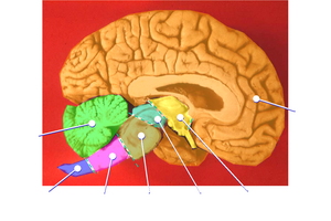
Human brain left midsagitttal view closeup description 2.JPG 701 × 490; 61 KB
-
Human brain left midsagitttal view closeup description 3.JPG 701 × 490; 58 KB
-
Human brainstem anterior view 2 description.JPG 346 × 487; 35 KB
-
Human brainstem anterior view description 2.JPG 347 × 485; 31 KB
-
Human brainstem anterior view description.JPG 347 × 485; 31 KB
-
Human brainstem Sagittal view.jpg 490 × 360; 146 KB
-
Hypofýza a pineální žláza.svg 114 × 76; 59 KB
-
Illu pituitary pineal glands ja.JPG 400 × 255; 23 KB
-
Illu pituitary pineal glands zh.jpg 400 × 259; 31 KB
-
Illu pituitary pineal glands-az.png 400 × 259; 84 KB
-
Illu pituitary pineal glands.jpg 400 × 259; 23 KB
-
Illu pituitary pineal glandsur.jpg 388 × 259; 39 KB
-
Location of the foramina of the fourth ventricle.jpg 650 × 462; 56 KB
-
Medulla oblongata, pons and middle cerebellar peduncle-es.png 1,000 × 750; 1.74 MB
-
Medulla oblongata, pons and middle cerebellar peduncle.jpg 960 × 720; 96 KB
-
Medulla oblongata, pons and middle cerebellar peduncle2.jpg 1,440 × 1,080; 308 KB
-
Midline sagittal view of the brainstem and cerebellum.png 688 × 505; 396 KB
-
Pons - high mag.jpg 4,272 × 2,848; 6.78 MB
-
Pons - intermed mag.jpg 4,272 × 2,848; 7.71 MB
-
Pons - very high mag.jpg 4,272 × 2,848; 6.27 MB
-
Pons and medulla oblongata.jpg 960 × 720; 112 KB
-
Pons image.png 800 × 455; 316 KB
-
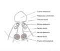
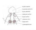
Pons section at facial colliculus-es.png 850 × 715; 165 KB
-
Pons section at facial colliculus.png 850 × 715; 136 KB
-
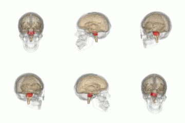
Pons small.gif 200 × 200; 563 KB
-
Rhombencephalon.jpg 960 × 720; 70 KB
-
Slide2cuc.JPG 960 × 720; 101 KB
-
Slide2MIR.JPG 960 × 720; 115 KB
-
Slide2PIT.JPG 960 × 720; 97 KB
-
Slide2RAFA-es.png 1,000 × 750; 768 KB
-
Slide2RAFA.JPG 960 × 720; 61 KB
-
Slide3MIR.JPG 960 × 720; 127 KB
-
Slide3PIT.JPG 960 × 720; 105 KB
-
Slide4MIR.JPG 960 × 720; 97 KB
-
Sobo 1909 648.png 1,063 × 1,048; 3.19 MB
-
Sobo 1909 656.png 867 × 498; 1.24 MB
-
Tentorum cerebelli.jpg 960 × 720; 81 KB
