Category:Human occipital bones
Jump to navigation
Jump to search
| Upload media | |||||
| Subclass of | |||||
|---|---|---|---|---|---|
| |||||
Subcategories
This category has the following 34 subcategories, out of 34 total.
3
A
B
- Basion (15 F)
C
- Cerebellar fossa (19 F)
- Cerebral fossa (15 F)
- Clivus (anatomy) (21 F)
- Condyloid fossa (18 F)
- Cruciform eminence (18 F)
E
- External occipital crest (9 F)
G
- Groove for transverse sinus (29 F)
H
I
- Inion (9 F)
- Internal occipital crest (19 F)
J
- Jugular process (36 F)
L
M
N
O
- Occipital condyles (42 F)
- Opisthion (18 F)
S
- Squama occipitalis (19 F)
Media in category "Human occipital bones"
The following 98 files are in this category, out of 98 total.
-
Basilar part.jpg 960 × 720; 109 KB
-
BodyParts3D FJ6411 Occipital bone.stl 5,120 × 2,880; 605 KB
-
Bones of the skull; four figures. Ink and watercolour, after Wellcome V0008218.jpg 3,129 × 2,330; 3.83 MB
-
Braus 1921 75.png 692 × 616; 1.22 MB
-
Building Fairs Brno 2011 (213).jpg 1,989 × 3,141; 3.42 MB
-
Condylar canal.jpg 708 × 518; 568 KB
-
Cranium - os occipitale (external).jpg 4,608 × 3,456; 4.69 MB
-
Cranium - os occipitale (internal).jpg 4,608 × 3,456; 4.05 MB
-
Cunningham’s Text-book of Anatomy (1914) - Fig 135.png 802 × 1,164; 1,009 KB
-
Cunningham’s Text-book of Anatomy (1914) - Fig 396.png 1,950 × 1,347; 1.86 MB
-
External occipital crest.jpg 960 × 720; 109 KB
-
External occipital protuberance.jpg 960 × 720; 107 KB
-
Gerrish's Text-book of Anatomy (1902) - Fig. 204.png 1,202 × 1,110; 997 KB
-
Gerrish's Text-book of Anatomy (1902) - Fig. 205.png 1,100 × 1,188; 1 MB
-
Gray 129 - Os Occipital - Surface-externe.png 550 × 529; 250 KB
-
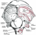
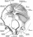
Gray 130 - Os Occipital - Surface-interne.png 535 × 600; 355 KB
-
Gray1031.png 550 × 659; 90 KB
-
Gray129.png 550 × 529; 55 KB
-
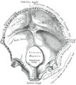
Gray130.png 535 × 600; 67 KB
-
Gray131.png 302 × 375; 25 KB
-
Gray187.png 718 × 1,169; 147 KB
-
Gray193.png 719 × 1,057; 150 KB
-
Gray194.png 650 × 420; 84 KB
-
Gray194az.png 650 × 420; 284 KB
-
Gray304.png 600 × 470; 60 KB
-
Gray307.png 600 × 416; 53 KB
-
Gray308.png 500 × 485; 51 KB
-
Gray387.png 443 × 600; 54 KB
-
Gray409.png 733 × 1,156; 172 KB
-
Gray70.png 600 × 597; 72 KB
-
Gray792.png 577 × 600; 96 KB
-
Gray800.png 600 × 382; 52 KB
-
Holden's human osteology (1899) - Plt05 Fig01.png 1,808 × 1,500; 2.02 MB
-
Holden's human osteology (1899) - Plt05 Fig02.png 1,803 × 1,563; 2.69 MB
-
Holden's human osteology (1899) - Plt21.png 1,678 × 2,805; 2.28 MB
-
Holden's human osteology (1899) - Plt45.png 1,431 × 1,020; 682 KB
-
Human occipital bone.stl 5,120 × 2,880; 4.8 MB
-
Internal occipital protuberance.jpg 960 × 720; 116 KB
-
Kort-lang-skalle.gif 3,072 × 1,883; 135 KB
-
Lambdoid suture.jpg 960 × 720; 62 KB
-
Lambdoid suture.png 1,600 × 1,200; 54 KB
-
Morris' human anatomy (1898) - Fig 030.png 1,624 × 992; 1.47 MB
-
Morris' human anatomy (1933) - Fig 140.png 2,216 × 1,904; 4.47 MB
-
Occipital (Holes Filled).stl 5,120 × 2,880; 21.53 MB
-
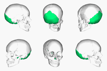
Occipital bone - animation 01.gif 600 × 600; 20 MB
-
Occipital bone - animation 02.gif 600 × 600; 21.24 MB
-
Occipital bone 090 000.png 600 × 600; 114 KB
-
Occipital bone 2.jpg 960 × 720; 87 KB
-
Occipital bone 3.jpg 960 × 720; 100 KB
-
Occipital bone 4.jpg 960 × 720; 110 KB
-
Occipital bone 5.jpg 960 × 720; 112 KB
-
Occipital bone animation.gif 320 × 320; 769 KB
-
Occipital bone animation2.gif 320 × 320; 1,014 KB
-
Occipital bone animation3.gif 320 × 320; 884 KB
-
Occipital bone close-up anterior.png 900 × 900; 203 KB
-
Occipital bone close-up inferior animation.gif 320 × 320; 846 KB
-
Occipital bone close-up inferior.png 900 × 900; 162 KB
-
Occipital bone close-up lateral animation.gif 320 × 320; 804 KB
-
Occipital bone close-up lateral.png 900 × 900; 107 KB
-
Occipital bone close-up suerperior.png 900 × 900; 229 KB
-
Occipital bone close-up suerperior2.png 900 × 900; 253 KB
-
Occipital bone close-up suerperior3.png 900 × 900; 248 KB
-
Occipital bone close-up superior animation.gif 320 × 320; 1 MB
-
Occipital bone inferior.png 900 × 900; 222 KB
-
Occipital bone inferior2.png 900 × 900; 229 KB
-
Occipital bone lateral.png 900 × 900; 144 KB
-
Occipital bone lateral2.png 900 × 900; 192 KB
-
Occipital bone lateral3.png 900 × 900; 159 KB
-
Occipital bone lateral4.png 4,500 × 4,500; 3.81 MB
-
Occipital bone lateral5.png 4,500 × 4,500; 3.86 MB
-
Occipital bone posterior.png 900 × 900; 150 KB
-
Occipital Bone Simple.png 600 × 600; 61 KB
-
Occipital bone suerperior.png 900 × 900; 96 KB
-
Occipital bone superior.png 900 × 900; 177 KB
-
Occipital bone.jpg 960 × 720; 76 KB
-
Occipital condyle.jpg 960 × 720; 103 KB
-
Occipital ridge.gif 2,070 × 2,214; 156 KB
-
OccipitalBun.jpg 222 × 231; 44 KB
-
Occipitomastoid suture.png 1,600 × 1,200; 52 KB
-
Piersol's human anatomy (1919) - Fig. 192.png 1,492 × 945; 1.09 MB
-
Piersol's human anatomy (1919) - Fig. 193.png 1,487 × 1,110; 1.54 MB
-
Piersol's human anatomy (1919) - Fig. 194.png 887 × 983; 669 KB
-
Posterior fossa.png 712 × 1,057; 497 KB
-
PSM V72 D309 Occiput of goat skull.png 834 × 1,011; 244 KB
-
Rotation Occipital bone.gif 600 × 600; 3.66 MB
-
SchaedelSeitlichSutur2.png 1,600 × 1,200; 32 KB
-
Skull and brainstem inner ear.svg 574 × 612; 702 KB
-
Skull base anatomy.jpg 1,847 × 2,952; 1.21 MB
-
Sobo 1909 106.png 1,364 × 1,516; 5.93 MB
-
Sobo 1909 243.png 2,100 × 1,820; 10.95 MB
-
Sobo 1909 47.png 1,660 × 2,020; 9.61 MB
-
Sobo 1909 48.png 2,256 × 1,528; 9.88 MB
-
Sobo 1909 49.png 2,240 × 1,680; 10.79 MB
-
Sobo 1909 50.png 1,860 × 1,140; 6.08 MB
-
Sobo 1909 51.png 2,084 × 2,148; 12.83 MB
-
Swanscombe occipital 01.jpg 1,818 × 1,344; 977 KB
-
Testut's Treatise on Human Anatomy (1911) - Vol 1 - Fig 152.png 777 × 1,275; 605 KB
-
WhiteDesertSkullCropped.png 496 × 381; 267 KB


