Category:Human maxilla
Jump to navigation
Jump to search
Subcategories
This category has the following 16 subcategories, out of 16 total.
3
- 3D data of human maxilla (1 F)
A
- Animations of human maxilla (13 F)
- Anterior nasal spine (17 F)
F
- Frontal process of maxilla (32 F)
H
I
- Infraorbital foramen (20 F)
M
O
P
- Palatine process of maxilla (31 F)
- Photographs of human maxilla (19 F)
R
Z
- Zygomatic process of maxilla (21 F)
- Zygomaticomaxillary sutures (27 F)
Media in category "Human maxilla"
The following 159 files are in this category, out of 159 total.
-
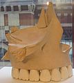
'Model of the Right Maxilla' by William Rush, c. 1808.JPG 2,175 × 2,440; 402 KB
-
711 Maxilla.jpg 784 × 601; 166 KB
-
-
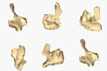
BodyParts3D FJ6380 FJ6468 Maxilla.stl 5,120 × 2,880; 510 KB
-
Cambridge Natural History Mammalia Fig 284.png 904 × 417; 50 KB
-
Canalis incisivus 18M - CT axial und sagittal - 001 - Annotation.jpg 1,524 × 838; 75 KB
-
Canalis incisivus 18M - CT axial und sagittal - 001.jpg 1,524 × 838; 75 KB
-
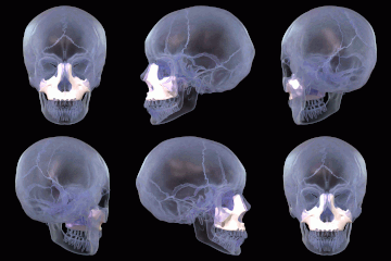
Cbct skull.jpg 538 × 614; 52 KB
-
Cranio. Norma laterale. Splancnocranio.jpg 960 × 720; 84 KB
-
Cranium - concha nasalis detail.jpg 4,608 × 3,456; 3.46 MB
-
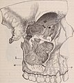
Cutaway image of human jaw with teeth (1917).jpg 529 × 493; 136 KB
-
Dental cosmos (1889) (14595327677).jpg 1,774 × 1,814; 421 KB
-
Dental cosmos (1889) (14781479972).jpg 1,588 × 1,924; 435 KB
-
Dental cosmos (1893) (14776794354).jpg 1,800 × 2,014; 383 KB
-
Dental cosmos (1893) (14779155935).jpg 1,396 × 2,162; 312 KB
-
Dixon's Manual of human osteology (1912) - Fig 108.png 1,306 × 1,114; 644 KB
-
Gerrish's Text-book of Anatomy (1902) - Fig. 222.png 1,304 × 828; 544 KB
-
Gerrish's Text-book of Anatomy (1902) - Fig. 223.png 1,260 × 890; 548 KB
-
Gerrish's Text-book of Anatomy (1902) - Fig. 224.png 858 × 934; 552 KB
-
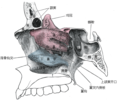
Gray153 zh.png 600 × 501; 200 KB
-
Gray153.png 600 × 501; 62 KB
-
Gray154.png 400 × 460; 50 KB
-
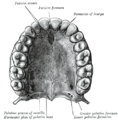
Gray157.png 600 × 434; 38 KB
-
Gray158.png 550 × 450; 38 KB
-
Gray159.png 700 × 503; 78 KB
-
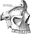
Gray160.png 492 × 500; 51 KB
-
Gray161.png 221 × 182; 5 KB
-
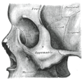
Gray162.png 185 × 189; 6 KB
-
Gray164 zh.png 400 × 379; 143 KB
-
Gray164-ar.png 400 × 379; 191 KB
-
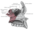
Gray164.png 400 × 379; 49 KB
-
Gray167.png 522 × 452; 41 KB
-
Gray173.az.png 550 × 487; 145 KB
-
Gray187.png 718 × 1,169; 147 KB
-
Gray191.png 500 × 465; 37 KB
-
Gray194.png 650 × 420; 84 KB
-
Gray196.png 600 × 428; 43 KB
-
Gray852.png 260 × 500; 29 KB
-
Gray853.png 400 × 329; 47 KB
-
Gray860.png 500 × 288; 25 KB
-
Gray861.png 600 × 358; 38 KB
-
Gray862.png 500 × 395; 39 KB
-
Gray995 zh.png 2,504 × 2,036; 2.31 MB
-
Gray995.png 2,504 × 2,036; 2.82 MB
-
Gray996.png 500 × 514; 50 KB
-
Holden's human osteology (1899) - Plt12 Fig01.png 1,696 × 1,194; 1.43 MB
-
Holden's human osteology (1899) - Plt12 Fig02.png 1,632 × 1,068; 1.28 MB
-
Holden's human osteology (1899) - Plt18 Fig01.png 1,460 × 1,992; 3.09 MB
-
Horizontal plate.jpg 960 × 720; 98 KB
-
Human skull - black and white.jpg 3,038 × 2,012; 1.82 MB
-
Human skull - close up.jpg 3,038 × 2,012; 2.97 MB
-
Human skull 2008.jpg 1,936 × 1,288; 1.71 MB
-
Humanjaw.png 334 × 401; 166 KB
-
Incisive fossa.jpg 960 × 720; 92 KB
-
Inferior viscerocranium.jpg 960 × 720; 81 KB
-
Jade-Toothed Skull.jpg 1,292 × 1,809; 2.13 MB
-
Jan25-skull.jpg 2,190 × 1,956; 440 KB
-
LeFort3 Osteotomie.png 1,600 × 1,200; 243 KB
-
Left maxilla close-up animation.gif 320 × 320; 970 KB
-
Left maxilla close-up anterior.png 900 × 900; 136 KB
-
Left maxilla close-up inferior animation.gif 320 × 320; 820 KB
-
Left maxilla close-up inferior.png 900 × 900; 155 KB
-
Left maxilla close-up lateral.png 900 × 900; 161 KB
-
Left maxilla close-up medial.png 900 × 900; 183 KB
-
Left maxilla close-up posterior.png 900 × 900; 149 KB
-
Left maxilla close-up superior animation.gif 320 × 320; 798 KB
-
Left maxilla close-up superior.png 900 × 900; 119 KB
-
Leprosy Cranium.JPG 1,536 × 2,304; 514 KB
-
Maastricht Sint-Servaasbasiliek BW 2017-08-19 10-57-27.jpg 6,016 × 4,000; 11.98 MB
-
Maastricht Sint-Servaasbasiliek BW 2017-08-19 10-57-40.jpg 6,016 × 4,000; 12.87 MB
-
Maxilla - animation 01.gif 600 × 600; 20.38 MB
-
Maxilla - animation 02.gif 600 × 600; 21.66 MB
-
Maxilla 1.jpg 960 × 720; 61 KB
-
Maxilla 2.jpg 960 × 720; 64 KB
-
Maxilla 4.jpg 960 × 720; 80 KB
-
Maxilla 5.jpg 960 × 720; 72 KB
-
Maxilla 6.jpg 960 × 720; 65 KB
-
Maxilla animation.gif 320 × 320; 916 KB
-
Maxilla anterior.png 900 × 900; 185 KB
-
Maxilla anterior2.png 900 × 900; 205 KB
-
Maxilla close-up animation.gif 320 × 320; 1.1 MB
-
Maxilla close-up anterior.png 900 × 900; 201 KB
-
Maxilla close-up inferior animation.gif 320 × 320; 1.15 MB
-
Maxilla close-up inferior.png 900 × 900; 178 KB
-
Maxilla close-up lateral.png 900 × 900; 140 KB
-
Maxilla close-up posterior.png 900 × 900; 228 KB
-
Maxilla close-up superior animation.gif 320 × 320; 1.16 MB
-
Maxilla close-up superior.png 900 × 900; 167 KB
-
Maxilla image.png 800 × 455; 307 KB
-
Maxilla inferior animation.gif 320 × 320; 1.04 MB
-
Maxilla inferior animation2.gif 320 × 320; 1.01 MB
-
Maxilla inferior.png 900 × 900; 206 KB
-
Maxilla inferior2.png 900 × 900; 215 KB
-
Maxilla inferior3.png 900 × 900; 220 KB
-
Maxilla kranial.png 728 × 477; 260 KB
-
Maxilla lateral (1).png 710 × 614; 414 KB
-
Maxilla lateral.png 900 × 900; 143 KB
-
Maxilla Simple.png 600 × 600; 61 KB
-
Maxilla superior animation.gif 320 × 320; 724 KB
-
Maxilla superior.png 900 × 900; 172 KB
-
Maxilla.jpg 960 × 720; 71 KB
-
Maxilla2.jpg 960 × 720; 65 KB
-
Maxilla3.jpg 960 × 720; 65 KB
-
Mid facelift (rhytidectomy) lower incision.png 1,152 × 966; 1.98 MB
-
Monte Albán - Gefeilte Zähne.jpg 2,560 × 1,920; 1.36 MB
-
Morris' human anatomy (1898) - Fig 084.png 1,548 × 1,912; 1.84 MB
-
Nasal bone.jpg 960 × 720; 65 KB
-
Nasenmuscheln1.JPG 3,168 × 2,376; 4.62 MB
-
Nosikaulis.jpg 200 × 230; 21 KB
-
Orbital bones.png 350 × 350; 104 KB
-
Orbital cavity.jpg 960 × 720; 98 KB
-
Orbite et Sinus maxillaire.png 735 × 608; 268 KB
-
Os maxillaire droit.jpg 765 × 567; 280 KB
-
Palate (PSF).png 3,240 × 1,384; 288 KB
-
Palate 1 (PSF).png 1,703 × 1,674; 459 KB
-
Palate 2 (PSF).png 1,341 × 1,238; 178 KB
-
Palatomaxilláris varrat.PNG 500 × 514; 51 KB
-
Piersol's human anatomy (1919) - Fig. 224.png 1,342 × 929; 1,009 KB
-
Piersol's human anatomy (1919) - Fig. 225.png 1,419 × 977; 832 KB
-
Piersol's human anatomy (1919) - Fig. 226.png 621 × 425; 151 KB
-
Piersol's human anatomy (1919) - Fig. 227.png 650 × 434; 151 KB
-
Pterygopalatine fossa.PNG 640 × 480; 743 KB
-
RhinoplastySkullOsteo.jpg 371 × 565; 36 KB
-
Rotation Maxilla.gif 600 × 600; 3.44 MB
-
SchaedelSeitlichSutur6.png 1,600 × 1,200; 32 KB
-
SchaedelSeitlichSutur8.png 1,600 × 1,200; 32 KB
-
SchaedelSeitlichSutur9.png 1,600 × 1,200; 32 KB
-
Siebert 12 (jaws).jpg 1,302 × 1,667; 558 KB
-
Siebert 12.jpg 1,946 × 2,854; 1.33 MB
-
Skull of Gough's Cave.jpg 2,671 × 2,025; 5.62 MB
-
Skullclose.jpg 853 × 1,280; 124 KB
-
Slide11hhhh.JPG 960 × 720; 83 KB
-
Slide12hhhh.JPG 960 × 720; 84 KB
-
Slide15aaa.JPG 960 × 720; 119 KB
-
Slide4fen.JPG 960 × 720; 115 KB
-
Slide8hhhh.JPG 960 × 720; 76 KB
-
Sobo 1906 338.png 1,512 × 1,419; 2.05 MB
-
Sobo 1909 100-es.png 1,471 × 1,179; 1.56 MB
-
Sobo 1909 100.png 1,476 × 1,184; 5.01 MB
-
Sobo 1909 101.png 2,068 × 1,588; 9.41 MB
-
Sobo 1909 102.png 1,928 × 1,628; 9 MB
-
Sobo 1909 103.png 1,372 × 1,100; 4.33 MB
-
Sobo 1909 76.png 1,492 × 1,316; 5.63 MB
-
Sobo 1909 77.png 1,732 × 1,224; 6.08 MB
-
Sobo 1909 79.png 1,480 × 1,248; 5.29 MB
-
Sobo 1909 80.png 1,336 × 1,080; 4.14 MB
-
Sobo 1909 81.png 1,248 × 1,152; 4.12 MB
-
Sobo 1909 96.png 1,720 × 1,616; 7.97 MB
-
Sobo 1909 98.png 2,308 × 1,476; 2.74 MB
-
Sobo 1909 99.png 1,628 × 1,196; 5.58 MB
-
Surface anatomy of maxillary denture-bearing area.png 1,018 × 554; 379 KB
-
Suturapalatinatransversum.PNG 492 × 500; 51 KB
-
Symphysis menti (Gray190 edit)-ar.svg 397 × 294; 599 KB
-
Teethsideview.jpg 2,944 × 2,468; 2.99 MB
-
Teethsideviewwp.jpg 2,944 × 2,468; 3.28 MB
-
Testut's Treatise on Human Anatomy (1911) - Vol 1 - Fig 189.png 879 × 915; 348 KB
-
Upper jaw.jpg 903 × 677; 303 KB
-
Virsutinis zandikaulis.JPG 1,140 × 780; 140 KB
-
Viršutinis žandikaulis, maxilla.jpg 1,140 × 780; 540 KB

