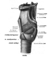
Category:Human larynx
Jump to navigation
Jump to search
Subcategories
This category has the following 13 subcategories, out of 13 total.
A
- Arytenoid cartilages (4 F)
C
- Cricoid cartilage (7 F)
D
E
- Epiglottis (1 P, 21 F)
G
H
L
- Laryngeal mirror (5 F)
S
T
- Thyroid cartilage (8 F)
U
V
Media in category "Human larynx"
The following 132 files are in this category, out of 132 total.
-
2306 The Larynx.jpg 1,814 × 1,673; 649 KB
-
Acute inflammation of the larynx Wellcome L0062698.jpg 4,795 × 6,173; 5.07 MB
-
An essay on the diseases of the larynx and trachea Wellcome L0029606.jpg 2,946 × 3,800; 3.42 MB
-
Anatomy, physiology and hygiene (1900) (14592657709).jpg 960 × 1,536; 525 KB
-
Anatomytool larynx and vocal cords English.jpg 555 × 556; 105 KB
-
Anesthetic-Management-for-Laser-Excision-of-Ball-Valving-Laryngeal-Masses-875053.f1.ogv 9.4 s, 568 × 320; 933 KB
-
Ausgepraegte Verkalkungen des Schildknorpels im Roentgenbild.jpg 1,036 × 1,402; 128 KB
-
Browne and Behnke, larynx, "Voice, song, and speech" Wellcome L0012859.jpg 1,152 × 1,606; 461 KB
-
Die Gartenlaube (1875) b 633 2.jpg 701 × 833; 227 KB
-
Doofstommen-onderwijs school in Amsterdam, SFA001001456 01.jpg 4,680 × 3,522; 1.29 MB
-
EB1911 Voice - action of the muscles of the larynx.jpg 957 × 885; 207 KB
-
EB1911 Voice - Cartilages and Ligaments of the Larynx (behind).jpg 334 × 477; 101 KB
-
EB1911 Voice - Cartilages and Ligaments of the Larynx (front).jpg 323 × 472; 100 KB
-
Education of Deaf-Mutes - A Manual for Teachers (1888) (14779983975).jpg 2,012 × 3,002; 627 KB
-
Figure 37 05 02.jpg 544 × 544; 126 KB
-
Gray1174 az.jpg 624 × 600; 83 KB
-
Gray1174.png 586 × 600; 59 KB
-
Gray1204 zh.png 511 × 400; 113 KB
-
Gray1204.png 511 × 400; 46 KB
-
Gray950 (cropped2).png 395 × 206; 9 KB
-
Gray950.png 400 × 800; 44 KB
-
Gray951 zh.png 497 × 600; 165 KB
-
Gray951-Cartilages larynx - Vue antérieure.png 497 × 600; 239 KB
-
Gray951.png 497 × 600; 63 KB
-
Gray951ja.png 497 × 600; 57 KB
-
Gray952-Cartilages larynx - vue postèrieure.png 441 × 600; 216 KB
-
Gray952.png 441 × 600; 57 KB
-
Gray955 zh.png 550 × 536; 160 KB
-
Gray955.png 550 × 536; 61 KB
-
Gray958.png 363 × 500; 40 KB
-
Gray959.png 404 × 600; 45 KB
-
Gray960 muscles of larynx ru.png 489 × 600; 77 KB
-
Gray960.png 489 × 600; 65 KB
-
Hand-book of physiology (1892) (14765020862).jpg 708 × 1,236; 195 KB
-
Human Larynx Model (50692885828).jpg 3,456 × 4,608; 2.35 MB
-
Human Larynx Model (50692886813).jpg 2,904 × 3,966; 1.92 MB
-
Human Larynx Model (50692887438).jpg 2,490 × 2,410; 1.1 MB
-
Illu07 larynx02.jpg 492 × 246; 42 KB
-
Illustration of a man's, woman's and child's larynx. Wellcome L0075050.jpg 4,102 × 6,442; 7.55 MB
-
Kehlkopf (Meyers).jpg 472 × 337; 56 KB
-
Kehlkopf Mensch.jpg 1,920 × 1,416; 598 KB
-
Kehlkopf Schema.png 816 × 1,100; 398 KB
-
-
-
Laryngeale Penetration - Schluck seitlich - Annotation.jpg 700 × 1,128; 60 KB
-
Laryngeale Penetration - Schluck seitlich.jpg 700 × 1,128; 53 KB
-
Larynx (top view)-es.png 1,400 × 1,261; 406 KB
-
Larynx (top view).jpg 1,400 × 1,261; 252 KB
-
Larynx and nearby structures-ar.jpg 1,050 × 1,428; 341 KB
-
Larynx and nearby structures.jpg 1,050 × 1,428; 145 KB
-
Larynx and Pharynx Anatomical Relationship.png 5,025 × 2,520; 1.26 MB
-
Larynx Anterolateral View Unlabeled.jpg 1,550 × 1,733; 196 KB
-
Larynx endo 2.jpg 478 × 295; 31 KB
-
Larynx external az.png 553 × 479; 83 KB
-
Larynx external en.svg 1,100 × 950; 40 KB
-
Larynx from a case of croup Wellcome L0062699.jpg 3,458 × 5,073; 3.63 MB
-
Larynx illustrations, 19th century Wellcome L0030210.jpg 1,688 × 1,269; 795 KB
-
Larynx normal.jpg 800 × 600; 174 KB
-
Larynx normal1a.jpg 1,258 × 944; 220 KB
-
Larynx normal2a.jpg 1,176 × 882; 182 KB
-
Larynx opened.jpg 960 × 720; 109 KB
-
Larynx PDW nima.jpg 1,024 × 768; 137 KB
-
Larynx Willis Gray 1858 681.png 1,189 × 1,480; 1.25 MB
-
Larynx, 17th century Wellcome L0007989.jpg 1,115 × 1,702; 818 KB
-
Larynx, 17th century Wellcome L0007990.jpg 1,046 × 1,840; 538 KB
-
Larynx.jpg 960 × 720; 99 KB
-
Larynx101.jpg 500 × 815; 110 KB
-
Larynxmikro2.jpg 1,445 × 1,084; 242 KB
-
Larynxmikro3.jpg 2,272 × 1,704; 564 KB
-
Macewan-type endotracheal tubes, United Kingdom, 1871-1900 Wellcome L0058070.jpg 4,398 × 3,222; 1.91 MB
-
Musculusaryepiglotticus.png 404 × 600; 46 KB
-
Musculusarytenoideus.png 363 × 500; 43 KB
-
Musculuscricoarytenoideuslateralis.png 404 × 600; 46 KB
-
Musculuscricoarytenoideusposterior.png 404 × 600; 46 KB
-
Musculuscricopharyngeus.PNG 400 × 665; 85 KB
-
Musculuscricothyreoideus.png 404 × 600; 46 KB
-
Musculusthyreoarytenoideus.png 489 × 600; 65 KB
-
Musculusthyroepiglotticus.png 404 × 600; 46 KB
-
Musculusuvulae.png 500 × 600; 322 KB
-
Normal Epiglottis.jpg 725 × 723; 37 KB
-
O'Dwyer-type intubation set, France, 1882-1900 Wellcome L0057986.jpg 4,112 × 2,832; 1.3 MB
-
Peroral endoscopy and laryngeal surgery (1915) (14593231840).jpg 1,210 × 960; 94 KB
-
Peroral endoscopy and laryngeal surgery (1915) (14799770113).jpg 928 × 1,340; 77 KB
-
PSM V64 D268 Vocal cords during inspiration.png 495 × 436; 39 KB
-
PSM V64 D268 Vocal cords in position for speaking or singing.png 499 × 415; 30 KB
-
PSM V64 D269 Vertical sections of the larynx.png 1,043 × 443; 33 KB
-
PSM V64 D269 Vocal chords during expiration.png 496 × 414; 35 KB
-
PSM V64 D272 Larynx examination.png 815 × 842; 80 KB
-
PSM V64 D273 Stroboscope for examining vocal chord vibration.png 849 × 1,211; 159 KB
-
-
-
Schema thyroide larynx az.png 718 × 799; 164 KB
-
Slide4ooo.JPG 960 × 720; 77 KB
-
Sobo 1906 433.png 1,293 × 1,452; 1.79 MB
-
Sobo 1906 434.png 1,410 × 1,530; 2.06 MB
-
Sobo 1906 439.png 1,386 × 1,371; 1.82 MB
-
Sobo 1906 440.png 1,593 × 1,536; 2.34 MB
-
Sobo 1906 441.png 1,191 × 972; 1.11 MB
-
Sobo 1906 442.png 1,260 × 1,461; 1.76 MB
-
Stimmlippengranulom1.jpg 1,717 × 1,288; 360 KB
-
Stroboscopy Normal Female Vocal Cords.webm 1 min 3 s, 1,280 × 720; 8.37 MB
-
The common frog (Page 77, Figs. 39-40) BHL7743550.jpg 2,099 × 3,308; 542 KB
-
The pathology of the membrane of the larynx and bronchia (1809) (14580378060).jpg 1,484 × 2,812; 673 KB
-
The principles and practice of surgery (1916) (14761841074).jpg 1,426 × 1,488; 271 KB
-
Thyroid cartilage.jpg 960 × 720; 111 KB
-
Thyroid2 ukrainian.png 586 × 600; 81 KB
-
Tobold-type laryngeal syringe, London, England, 1902-1930 Wellcome L0058071.jpg 2,832 × 4,256; 1.24 MB
-
Vibrante simple vs Vibrante multiple en español.png 830 × 578; 201 KB
-
Vocal cord granuloma.jpg 900 × 676; 52 KB
-
Vocal folds-201611.jpg 639 × 474; 71 KB
-
Vocal folds-speaking 201611.jpg 511 × 375; 50 KB
-
Vorlesungen über die Krankheiten des Kehlkopfes (1893) (14591603468).jpg 1,504 × 3,348; 704 KB
-
Vorlesungen über die Krankheiten des Kehlkopfes (1893) (14591829887).jpg 1,964 × 3,352; 1.48 MB
-
Vorlesungen über die Krankheiten des Kehlkopfes (1893) (14775075341).jpg 1,536 × 3,732; 1.31 MB
-
Vorlesungen über die Krankheiten des Kehlkopfes (1893) (14777838862).jpg 2,076 × 3,216; 764 KB
-
Vorlesungen über die Krankheiten des Kehlkopfes (1893) (14777843512).jpg 1,304 × 2,268; 724 KB
-
Vorlesungen über die Krankheiten des Kehlkopfes (1893) (14798047223).jpg 2,372 × 2,816; 607 KB
-
Whistler-type laryngeal cutting dilator, London, England, 19 Wellcome L0058072.jpg 4,256 × 2,832; 1.01 MB
-
喉頭俯視圖.gif 369 × 270; 53 KB
-
喉頭正面圖.jpg 500 × 700; 48 KB


