Category:Human histology
Jump to navigation
Jump to search
Subcategories
This category has the following 14 subcategories, out of 14 total.
Media in category "Human histology"
The following 67 files are in this category, out of 67 total.
-
2UT-SAC-PDA.jpg 406 × 342; 136 KB
-
Acinar cells.JPG 2,816 × 2,112; 2.75 MB
-
Adiposo.jpg 1,289 × 1,519; 1.11 MB
-
ARTERIA1.JPG 1,600 × 1,200; 333 KB
-
Atlas and epitome of gynecology (1900) (14760773541).jpg 2,508 × 4,080; 2.5 MB
-
Ca in situ, cervix.jpg 1,818 × 1,228; 999 KB
-
Capsule of the Human Thyroid Gland (32534056467).jpg 3,264 × 1,840; 1.46 MB
-
Cell surface.jpg 1,382 × 1,036; 645 KB
-
Cementocitos.jpg 86 × 112; 2 KB
-
Colloid Filled Lumens in Human Thyroid Gland (32546734627).jpg 3,264 × 1,840; 5.65 MB
-
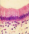
COLUMNAR SIMPLE LM.jpg 304 × 355; 56 KB
-
Cuboidal Epithelium Lining Intralobular Duct by Phase Contrast (33859250438).jpg 3,264 × 1,840; 6.52 MB
-
Deeply Septated Lobes of the Human Thyroid Gland (32534056797).jpg 3,264 × 1,840; 1.55 MB
-
Deeply Septated Lobes of the Human Thyroid Gland (32546733667).jpg 3,264 × 1,840; 6.72 MB
-
Deeply Septated Lobes of the Human Thyroid Gland by Phase Contrast (40523036103).jpg 3,264 × 1,840; 1.66 MB
-
Dense regular1.jpg 2,048 × 1,021; 459 KB
-
Dense regular2.jpg 1,881 × 1,077; 434 KB
-
Dense regular3.jpg 1,917 × 1,069; 445 KB
-
Dense regular4.jpg 1,886 × 1,205; 462 KB
-
Dysplastic gangliocytoma lhermitte duclos.jpg 2,080 × 1,542; 1.04 MB
-
Enamel and dentine - ground section.jpg 600 × 800; 178 KB
-
Ens th.svg 283 × 251; 99 KB
-
Epith simple columnar1.jpg 2,048 × 1,536; 736 KB
-
Epith simple columnar2.jpg 2,048 × 1,536; 736 KB
-
Epith simple columnar3.jpg 2,048 × 1,536; 772 KB
-
Epith simple columnar4.jpg 2,048 × 1,536; 743 KB
-
Epith simple columnar5.jpg 2,048 × 1,536; 736 KB
-
Epith simple cuboidal3.jpg 1,475 × 594; 198 KB
-
Epith simple cuboidal4.jpg 2,048 × 1,536; 654 KB
-
Epith simple cuboidal5.jpg 2,048 × 1,165; 520 KB
-
Epithelial Tissues Simple Squamous Epithelium (40823230315).jpg 3,264 × 1,840; 1.67 MB
-
Epithelial Tissues Simple Squamous Epithelium (41722161021).jpg 3,264 × 1,840; 1.51 MB
-
Epithelial Tissues Simple Squamous Epithelium (41722161301).jpg 3,264 × 1,840; 1.15 MB
-
Fibroso.jpg 1,289 × 1,519; 1.07 MB
-
Hassal.jpg 1,321 × 1,167; 601 KB
-
Histologie prostaty.png 600 × 874; 762 KB
-
Human brain disorganized by freezing. Wellcome L0003408.jpg 1,232 × 1,702; 986 KB
-
Human Simple Squamous Epithelium Tissue.jpg 1,796 × 2,006; 1.01 MB
-
Human tonsils (26 2 05) Cross-section; embedded in Epon.jpg 2,776 × 1,777; 629 KB
-
Humanskinsweatglands100x1.jpg 1,024 × 768; 170 KB
-
Humanskinsweatglands400x6.jpg 1,024 × 768; 137 KB
-
Humanskinsweatglands40x.jpg 1,024 × 768; 159 KB
-
Humanskinsweatglands40x8.jpg 1,024 × 768; 217 KB
-
Intercalated disc.png 450 × 295; 60 KB
-
INTESTINO 2.JPG 1,600 × 1,200; 389 KB
-
Intestino aaas.JPG 1,434 × 1,200; 362 KB
-
Kidney-Cortex.JPG 2,816 × 2,112; 2.21 MB
-
Large vein-Inferior vena cava.jpg 1,024 × 768; 88 KB
-
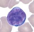
Lymphocyte GL.jpg 417 × 393; 17 KB
-
Migration routes of modern humans (2023).png 2,560 × 1,424; 693 KB
-
Oesophagus.jpg 800 × 704; 126 KB
-
Oviduct-histo.jpg 781 × 540; 156 KB
-
Peripheral nerve, cross section.jpg 709 × 532; 89 KB
-
Pinealocytes and Astrocytes in Older Human Pituitary by Phase Contrast (46684388614).jpg 3,264 × 1,840; 1.48 MB
-
Principal types of mankind.jpg 3,256 × 2,512; 1.34 MB
-
Scalloped Colloid in Follicles of the Human Thyroid Gland (47435785862).jpg 3,264 × 1,840; 5.8 MB
-
Stomaco.JPG 1,348 × 1,030; 378 KB
-
Striato.JPG 1,600 × 1,200; 510 KB
-
Tessuto nervoso 4.JPG 3,296 × 2,472; 2.01 MB
-
Tessuto nervoso 5.JPG 3,296 × 2,472; 1.89 MB
-
Tessuto nervoso 6.JPG 3,296 × 2,472; 1.94 MB
-
Tessuto nervoso2.JPG 3,296 × 2,472; 1.65 MB
-
Tessuto nervoso3.JPG 3,296 × 2,472; 1.97 MB
-
Ureter.JPG 2,816 × 2,112; 2.79 MB
-
Vascular Supply to the Septa of the Human Thyroid Gland (32534056677).jpg 3,264 × 1,840; 1.53 MB
-
Yellow adipose tissue in paraffin section - lipids washed out.jpg 746 × 535; 94 KB


