Category:Human heart cross-section
Jump to navigation
Jump to search
Subcategories
This category has only the following subcategory.
S
- SVG human heart cross-section (106 F)
Media in category "Human heart cross-section"
The following 135 files are in this category, out of 135 total.
-
2008 Internal Anatomy of the HeartN.jpg 1,656 × 1,159; 760 KB
-
201405 heart.png 400 × 400; 70 KB
-
2018 Conduction System of Heart.jpg 2,021 × 1,354; 1.03 MB
-
Anatomical plate of the heart (no labels).png 800 × 590; 230 KB
-
Aorta.jpg 317 × 462; 46 KB
-
ApCORAZON.png 225 × 252; 48 KB
-
Auricle (PSF).png 2,603 × 2,510; 333 KB
-
Blausen 0046 ArterialSwitchOperation 01.png 378 × 945; 351 KB
-
Blausen 0047 ArterialSwitchOperation 02.png 640 × 480; 344 KB
-
Blausen 0100 Bradycardia Fainting.png 640 × 480; 260 KB
-
Blausen 0452 Heart BloodFlow.png 768 × 1,024; 435 KB
-
Blausen 0457 Heart SectionalAnatomy.png 2,200 × 2,200; 2.22 MB
-
Blausen 0462 HeartAnatomy-es.png 768 × 1,024; 144 KB
-
Blausen 0462 HeartAnatomy.png 768 × 1,024; 461 KB
-
Blood Circulation.gif 700 × 650; 466 KB
-
Bundleofhis.png 400 × 483; 69 KB
-
Capasdelcorazon.png 947 × 1,465; 2.82 MB
-
Cardiac-Cycle-Animated.gif 1,024 × 1,536; 6.11 MB
-
CG Heart (cropped).gif 434 × 513; 3.19 MB
-
CG heart 2.gif 593 × 701; 6.78 MB
-
CG heart 90fps.webm 29 s, 570 × 678; 57.49 MB
-
CG Heart.gif 800 × 600; 3.27 MB
-
Ch19 heart hollow.png 784 × 892; 133 KB
-
CIRCULATION Heart Section.jpg 1,034 × 1,092; 268 KB
-
Coao.jpg 600 × 630; 88 KB
-
Cor -LFN.png 640 × 476; 151 KB
-
Corazón - Válvulas - 001.jpg 1,024 × 768; 281 KB
-
D-TGA.jpg 1,000 × 600; 457 KB
-
De-Geleidingssysteem (CardioNetworks ECGpedia).jpg 550 × 600; 42 KB
-
Der Herzschlag.jpg 3,507 × 2,550; 822 KB
-
Diagram of the human heart (catalan).png 600 × 600; 113 KB
-
Diagram of the human heart (cropped) az.png 600 × 600; 118 KB
-
Diagram of the human heart (cropped) el.png 600 × 600; 100 KB
-
Diagram of the human heart (cropped)(ZH T).png 612 × 624; 143 KB
-
Diagram of the human heart (cropped)-it.png 814 × 823; 173 KB
-
Diagram of the Human Heart.jpg 1,503 × 1,183; 683 KB
-
DiagramaCorazón.png 464 × 454; 99 KB
-
Echo heart parasternal long axis (CardioNetworks ECHOpedia).jpg 1,200 × 904; 1.11 MB
-
El corazón humano - Laura Macías Álvarez.jpg 1,169 × 1,654; 1.06 MB
-
Geleidingssysteem (CardioNetworks ECGpedia).jpg 800 × 872; 99 KB
-
Gray501.png 400 × 483; 65 KB
-
Grierson 15 Corazón.JPG 819 × 908; 163 KB
-
Hartas2 (CardioNetworks ECGpedia).jpg 800 × 793; 39 KB
-
Heart (CardioNetworks ECGpedia).svg 575 × 516; 38 KB
-
Heart anatomy in armenian..jpg 584 × 600; 70 KB
-
Heart anterior view coronal section.jpg 947 × 1,465; 1.3 MB
-
Heart aortic short axis section (CardioNetworks ECHOpedia).jpg 2,000 × 2,000; 950 KB
-
Heart aortic short axis section.jpg 2,000 × 2,000; 954 KB
-
Heart apical 2C anatomy.jpg 2,100 × 1,562; 1.34 MB
-
Heart apical 2chamber (CardioNetworks ECHOpedia).jpg 1,024 × 1,617; 228 KB
-
Heart apical 2chamber.jpg 1,133 × 1,789; 991 KB
-
Heart apical 4c anatomy (CardioNetworks ECHOpedia).jpg 512 × 691; 76 KB
-
Heart apical 4c anatomy.jpg 1,556 × 2,100; 1.52 MB
-
Heart coronal xs.jpg 1,475 × 972; 1.44 MB
-
Heart Diagram (PSF).png 3,247 × 2,689; 281 KB
-
Heart diagram corrected labels.JPG 569 × 386; 34 KB
-
Heart diagram-fa.PNG 839 × 655; 238 KB
-
Heart diastole.png 155 × 200; 9 KB
-
Heart labelled large prevedeno.PNG 530 × 526; 36 KB
-
Heart labelled large zh-cn.jpg 661 × 652; 53 KB
-
Heart labelled large.png 524 × 526; 36 KB
-
Heart left anterior oblique xs.jpg 957 × 1,483; 1.13 MB
-
Heart left atrial appendage tee view.jpg 1,133 × 1,789; 930 KB
-
Heart left lateral view.jpg 900 × 959; 951 KB
-
Heart left parasternal long axis.jpg 3,000 × 2,000; 1.19 MB
-
Heart left ventricular outflow track.jpg 1,720 × 2,184; 1.06 MB
-
Heart lpla echo view.jpg 3,000 × 2,000; 1.28 MB
-
Heart lpla echocardiography diagram.jpg 1,200 × 904; 1.15 MB
-
Heart mediastinum coronal xs.jpg 1,924 × 2,988; 1.77 MB
-
Heart normal short axis echo (CardioNetworks ECHOpedia).png 1,000 × 718; 31 KB
-
Heart normal short axis echo (CardioNetworks ECHOpedia).svg 480 × 344; 20 KB
-
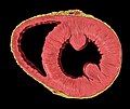
Heart normal short axis section.jpg 1,668 × 1,396; 1.05 MB
-
Heart Normal vs. Abnormal Flow To Lungs.png 400 × 1,270; 498 KB
-
Heart Normal vs. Enlarged.png 338 × 927; 919 KB
-
Heart numlabels.png 957 × 965; 237 KB
-
Heart right anatomy.jpg 2,179 × 2,108; 1.25 MB
-
Heart right asd.jpg 2,179 × 2,108; 1.24 MB
-
Heart right lateral view.jpg 1,313 × 967; 1.57 MB
-
Heart right vsd.jpg 2,179 × 2,108; 1.23 MB
-
Heart short axis echocardiography view.jpg 952 × 1,312; 756 KB
-
Heart short axis transgastric view.jpg 1,829 × 1,537; 996 KB
-
Heart short axis view papillary.jpg 952 × 1,312; 748 KB
-
Heart subcostal echocardiography view.jpg 1,884 × 2,211; 1.01 MB
-
Heart systole.png 155 × 200; 9 KB
-
Heart tee four chamber view.jpg 1,981 × 1,555; 1.48 MB
-
Heart tee tricuspid valve.jpg 1,841 × 1,677; 888 KB
-
Heart valves (heart schematic).png 696 × 503; 80 KB
-
Heart ventricular aneurysm views.jpg 459 × 393; 166 KB
-
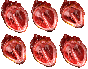
Heart X-sec.png 300 × 424; 373 KB
-
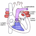
Herz Lungenkreislauf.png 957 × 965; 194 KB
-
Herz Schema.jpg 800 × 363; 168 KB
-
Herz-Heart.jpg 700 × 487; 98 KB
-
Human Heart.jpg 893 × 967; 394 KB
-
Hypoplastic Left Heart Syndrome.png 300 × 549; 182 KB
-
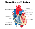
Ihmisen sydän poikkileikkaus.svg 981 × 817; 98 KB
-
Illu systemic circuit.jpg 300 × 500; 31 KB
-
Latidos.gif 300 × 300; 54 KB
-
Left atrial enlargement (CardioNetworks ECGpedia).jpg 800 × 843; 82 KB
-
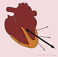
Left axis dev (CardioNetworks ECGpedia).jpg 798 × 819; 42 KB
-
Mapa do coração.PNG 758 × 586; 186 KB
-
Normal heart.jpg 650 × 600; 282 KB
-
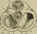
Orifices of the Heart seen from above in An academic physiology and hygiene (1903).jpg 1,612 × 1,472; 414 KB
-
Pulmonary artery catheter.png 488 × 680; 59 KB
-
Pulmonary Circuit.gif 450 × 332; 45 KB
-
Relations of the aorta, trachea, esophagus and other heart structures esp.png 2,089 × 1,645; 13.13 MB
-
Relations of the aorta, trachea, esophagus and other heart structures vi.png 2,089 × 1,645; 1.09 MB
-
Relations of the aorta, trachea, esophagus and other heart structures.png 2,089 × 1,645; 1.06 MB
-
Schéma srdce.svg 451 × 310; 55 KB
-
Section through heart to show valves and blood flow.jpg 602 × 454; 59 KB
-
Skilvelių pertvaros defektas.jpg 982 × 660; 98 KB
-
Sobo 1906 520.png 1,371 × 1,797; 7.06 MB
-
Sobo 1906 521.png 1,447 × 1,767; 7.33 MB
-
Sobo 1906 522.png 1,296 × 1,617; 6.01 MB
-
Sobo 1906 523.png 1,639 × 1,584; 7.44 MB
-
Sobo 1906 528.png 1,482 × 1,668; 729 KB
-
Sobo 1906 529.png 1,344 × 1,581; 2.03 MB
-
Stamp of Indonesia - 1972 - Colnect 257424 - World Heart Day.jpeg 269 × 358; 34 KB
-
TGV - schéma.gif 307 × 405; 21 KB
-
Vesalius, De humani corporis fabrica, 1543 Wellcome L0028440.jpg 1,667 × 1,288; 948 KB
-
Vidinė širdies sandara 1.png 658 × 472; 194 KB
-
W. Cowper, Myotomia reformata Wellcome L0024328.jpg 1,184 × 1,572; 823 KB
-
Šidis ir jos dalys.png 800 × 600; 377 KB
-
Širdies anatominė sandara.png 1,570 × 1,096; 804 KB
-
Širdies laidžioji sistema.png 1,041 × 768; 411 KB
-
Širdies sandara Pav 2.png 461 × 331; 127 KB
-
Širdies sandara.jpg 245 × 205; 12 KB
-
Širdis.png 799 × 646; 529 KB
-
Рейля канатик.jpg 228 × 210; 60 KB
-
Сердце.jpg 370 × 450; 17 KB
-
拡張型心筋症.jpg 960 × 720; 55 KB
-
肥大型心筋症.jpg 960 × 720; 53 KB
-
심장 수정.png 627 × 711; 453 KB


