Category:Human frontal bones
Jump to navigation
Jump to search
Subcategories
This category has the following 20 subcategories, out of 20 total.
3
A
B
C
F
G
- Gawis cranium (2 F)
- Glabella (anatomy) (5 F)
H
N
- Nasion (14 F)
O
P
S
T
Z
- Zygomaticofrontal sutures (10 F)
Media in category "Human frontal bones"
The following 124 files are in this category, out of 124 total.
-
De-Stirnbein.ogg 1.8 s; 20 KB
-
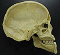
 Anthroplogy - human skull of a boy.JPG 4,608 × 3,072; 7.71 MB
-
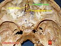
 Base du crane.png 946 × 1,284; 388 KB
-
 Base of skull 1.jpg 960 × 720; 116 KB
-
 BCH-808-Syphillis-Lo.jpg 2,624 × 2,656; 527 KB
-
 Bidloo Ontleding 1690 89.jpg 1,200 × 1,612; 242 KB
-
 BodyParts3D FJ6310 Frontal bone.stl 5,120 × 2,880; 706 KB
-
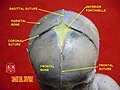
 Coronal suture.jpg 960 × 720; 65 KB
-
 Crane suture metopique 01 04 2012 2 B.jpg 1,985 × 1,624; 2.38 MB
-
 Cranio. Norma laterale. Splancnocranio.jpg 960 × 720; 84 KB
-
 Cranium - os ethmoidale, lamina cribrosa (superior view, detail).jpg 4,608 × 3,456; 3.45 MB
-
 Cunningham’s Text-book of Anatomy (1914) - Fig 130.png 806 × 804; 860 KB
-
 Demonstration model of the human eye, French. 1884 Wellcome S0008222.jpg 1,525 × 1,268; 749 KB
-
 Dolichocephalie.jpg 269 × 379; 27 KB
-
 Déformation Péruvienne MHNT Noir.jpg 5,271 × 5,023; 11.69 MB
-
 Eminentiafrontalis.PNG 518 × 500; 39 KB
-
 Forehead bulk defect picture 1.jpg 886 × 1,323; 245 KB
-
 Fraktur Stirnhoehle VR.png 1,080 × 684; 974 KB
-
 Frauenschädel Regensburg-Harting.jpg 1,643 × 2,191; 1.52 MB
-
 Frontal Bone (Holes Filled).stl 5,120 × 2,880; 41.13 MB
-
 Frontal bone - animation 01.gif 600 × 600; 17.02 MB
-
 Frontal bone - animation 02.gif 600 × 600; 20.43 MB
-
 Frontal bone animation.gif 320 × 320; 879 KB
-
 Frontal bone animation2.gif 320 × 320; 944 KB
-
 Frontal bone animation3.gif 320 × 320; 646 KB
-
 Frontal bone anterior.png 900 × 900; 194 KB
-
 Frontal bone anterior2.png 900 × 900; 167 KB
-
 Frontal bone close-up animation.gif 320 × 320; 1,008 KB
-
 Frontal bone close-up anterior.png 900 × 900; 187 KB
-
 Frontal bone close-up inferior animation.gif 320 × 320; 1.1 MB
-
 Frontal bone close-up inferior.png 900 × 900; 216 KB
-
 Frontal bone close-up inferior2.png 900 × 900; 186 KB
-
 Frontal bone close-up inferior3.png 900 × 900; 256 KB
-
 Frontal bone close-up lateral.png 900 × 900; 135 KB
-
 Frontal bone close-up posterior.png 900 × 900; 295 KB
-
 Frontal bone close-up superior.png 900 × 900; 130 KB
-
 Frontal bone hrtawiki.png 1,455 × 1,204; 1.08 MB
-
 Frontal bone inferior.png 900 × 900; 189 KB
-
 Frontal bone lateral.png 900 × 900; 149 KB
-
 Frontal bone lateral2.png 900 × 900; 175 KB
-
 Frontal bone lateral3.png 4,500 × 4,500; 1.58 MB
-
 Frontal Bone Simple.png 600 × 600; 59 KB
-
 Frontal bone superior.png 900 × 900; 109 KB
-
 Frontal bone superior2.png 900 × 900; 186 KB
-
 Frontal bone superior3.png 900 × 900; 160 KB
-
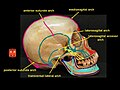
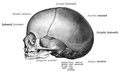
 Frontal bone.jpg 960 × 720; 86 KB
-
 Frontal bone.png 800 × 455; 243 KB
-
 Frontal suture.jpg 960 × 720; 65 KB
-
 Frontal.png 660 × 642; 166 KB
-
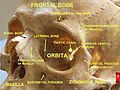
 Functional architecture of cranium 2.jpg 960 × 720; 77 KB
-
 Functional architecture of cranium.jpg 960 × 720; 104 KB
-
 Gerrish's Text-book of Anatomy (1902) - Fig. 208.png 1,562 × 1,038; 857 KB
-
 Gerrish's Text-book of Anatomy (1902) - Fig. 209.png 1,260 × 1,148; 1.27 MB
-
 Gerrish's Text-book of Anatomy (1902) - Fig. 210.png 1,384 × 580; 566 KB
-
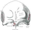 Gray134.png 518 × 500; 38 KB
-
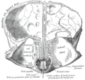 Gray135.png 550 × 518; 64 KB
-
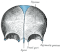 Gray136.png 400 × 343; 27 KB
-
 Gray159.png 700 × 503; 78 KB
-
 Gray164 zh.png 400 × 379; 143 KB
-
 Gray164-ar.png 400 × 379; 191 KB
-
 Gray164.png 400 × 379; 49 KB
-
 Gray193.png 719 × 1,057; 150 KB
-
 Gray194 zh.png 650 × 420; 245 KB
-
 Gray194.png 650 × 420; 84 KB
-
 Gray194az.png 650 × 420; 284 KB
-
 Gray379.png 535 × 450; 83 KB
-
 Gunshot skull civil war.jpg 880 × 1,200; 135 KB
-
 Holden's human osteology (1899) - Plt07 Fig01.png 1,590 × 1,431; 2.6 MB
-
 Holden's human osteology (1899) - Plt07 Fig02.png 1,686 × 1,488; 2.99 MB
-
 Holden's human osteology (1899) - Plt18 Fig01.png 1,460 × 1,992; 3.09 MB
-
 Human skull - close up.jpg 3,038 × 2,012; 2.97 MB
-
 Incan brain surgery.jpg 2,304 × 3,072; 1.18 MB
-
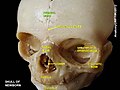
 Jan25-skull.jpg 2,190 × 1,956; 440 KB
-
 Kaktikaulis2.gif 867 × 569; 73 KB
-
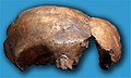
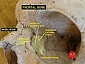
 KNM-ER 42700-Homo erectus.jpg 2,048 × 1,360; 1.2 MB
-
 Kort-lang-skalle.gif 3,072 × 1,883; 135 KB
-
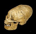
 La Carigüela-Neanderthal.jpg 1,305 × 783; 371 KB
-
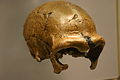 La Quina. H 5. Homo neanderthalensis.jpg 2,048 × 1,360; 1.3 MB
-
 Lacrimal and nasal bones.jpg 960 × 720; 86 KB
-
 MNH - Kauterisation.jpg 2,694 × 2,928; 1.14 MB
-
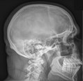
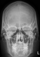 NNH im Roentgen frontal.png 678 × 962; 393 KB
-
 Orbital bones.png 350 × 350; 104 KB
-
 Orbital cavity.jpg 960 × 720; 98 KB
-
 Orbite et Sinus maxillaire.png 735 × 608; 268 KB
-
 Os frontal1.png 692 × 745; 255 KB
-
 Piece of skull showing a trephination experiment using a sha Wellcome L0058785.jpg 4,032 × 2,832; 1.19 MB
-
 Piersol's human anatomy (1919) - Fig. 220.png 1,488 × 1,226; 1.1 MB
-
 Piersol's human anatomy (1919) - Fig. 221.png 1,580 × 1,286; 1.7 MB
-

-
 Restos de Homo sapiens. Museu de Prehistòria de València 02.jpg 3,072 × 4,608; 5.48 MB
-
 Rotation frontal bone.gif 600 × 600; 3.18 MB
-
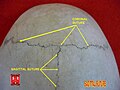 Sagittal suture 3.jpg 960 × 720; 60 KB
-
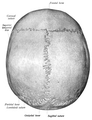
 Sagittal suture.jpg 960 × 720; 71 KB
-
 Schaedel im Röntgen seitlich.png 1,092 × 1,078; 640 KB
-
 SchaedelSeitlichSutur1.png 1,600 × 1,200; 32 KB
-
 SchaedelSeitlichSutur11.png 1,600 × 1,200; 32 KB
-
 SchaedelSeitlichSutur5.png 1,600 × 1,200; 32 KB
-
 SchaedelSeitlichSutur7.png 1,600 × 1,200; 32 KB
-
 SchaedelSeitlichSutur9.png 1,600 × 1,200; 32 KB
-
 Schädel Hahnöfersand front.jpg 2,048 × 1,990; 1.09 MB
-
 Schädel Hahnöfersand.jpg 2,048 × 1,190; 609 KB
-
 Skeleton with phrenological skull, Europe Wellcome L0057109.jpg 4,846 × 3,634; 3.96 MB
-
 Skull - midsaggital section P.2005.jpg 1,024 × 937; 62 KB
-
 Skull of Alaskan Inuit, effects of syphilis, 1910 Wellcome V0031332.jpg 2,161 × 3,228; 1.72 MB
-
 Skull sword trauma.jpg 1,200 × 1,022; 174 KB
-
 Skullfront.png 1,220 × 1,872; 1.81 MB
-
 Slide1nnnn.JPG 960 × 720; 57 KB
-
 Slide1pppp.JPG 960 × 720; 81 KB
-
 Slide27hhhh.JPG 960 × 720; 87 KB
-
 Slide3jjjj.JPG 960 × 720; 123 KB
-
 Sobo 1909 104.png 2,136 × 1,288; 7.89 MB
-
 Sobo 1909 46.png 1,656 × 2,168; 10.29 MB
-
 Sobo 1909 47.png 1,660 × 2,020; 9.61 MB
-
 Sobo 1909 66.png 2,228 × 1,380; 8.81 MB
-
 Sobo 1909 67.png 2,208 × 1,492; 9.44 MB
-
 Sobo 1909 68.png 1,948 × 1,016; 5.67 MB
-
 Sphenoid and temporal bones 2.jpg 960 × 720; 88 KB
-
 Sphenoid and temporal bones 3.jpg 960 × 720; 88 KB
-
 Sphenoid and temporal bones 4.jpg 960 × 720; 94 KB
-
 Superior viscerocranium.jpg 960 × 720; 76 KB
-
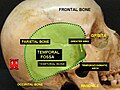 Temporal fossa.jpg 960 × 720; 87 KB
-
 Testut's Treatise on Human Anatomy (1911) - Vol 1 - Fig 128.png 662 × 852; 165 KB
-
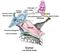 Vomer.png 635 × 572; 143 KB
-
 William Cheselden skull.jpg 884 × 1,456; 323 KB


.jpg)
_-_Fig_130.png)


_-_Fig._208.png)
_-_Fig._209.png)
_-_Fig._210.png)


_-_Plt07_Fig01.png)
_-_Plt07_Fig02.png)
_-_Plt18_Fig01.png)


_-_Fig._220.png)
_-_Fig._221.png)


_-_Vol_1_-_Fig_128.png)

