Category:Human embryology
Jump to navigation
Jump to search
Subcategories
This category has the following 10 subcategories, out of 10 total.
Media in category "Human embryology"
The following 93 files are in this category, out of 93 total.
-
2904 Preembryonic Development-02.jpg 1,957 × 1,792; 1.12 MB
-
2905 Implantation.jpg 1,950 × 2,440; 1.04 MB
-
2907 Embroyonic Disc, Amniotic Cavity, Yolk Sac-02-NLtxt.jpg 1,503 × 1,055; 689 KB
-
2907 Embroyonic Disc, Amniotic Cavity, Yolk Sac-02.jpg 1,529 × 1,055; 551 KB
-
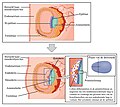
2908 Germ Layers-02-nltxt.jpg 1,854 × 1,658; 1.45 MB
-
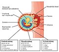
2908 Germ Layers-02.jpg 1,781 × 1,590; 1.06 MB
-
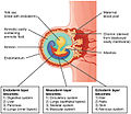
2909 Embryo Week 3-02 NLtxt.jpg 1,446 × 1,276; 855 KB
-
2909 Embryo Week 3-02.jpg 1,447 × 1,252; 634 KB
-
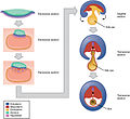
2913 Embryonic Folding.jpg 1,894 × 1,735; 831 KB
-
A textbook of obstetrics (1898) (14594028468).jpg 4,160 × 2,535; 836 KB
-
Development of human embryo at five stages. Wellcome L0057765.jpg 2,894 × 3,680; 1.53 MB
-
Development of human embryo at five stages. Wellcome L0057766.jpg 2,835 × 3,877; 1.2 MB
-
Development of human embryo at seven stages. Wellcome L0057764.jpg 2,835 × 3,866; 1.28 MB
-
Embryonalentwicklung (Jakob Ruf).png 282 × 204; 119 KB
-
Fertilization.jpg 1,920 × 1,080; 1.35 MB
-
Foetus in placenta in utero. Wellcome L0057759.jpg 2,953 × 3,713; 1.33 MB
-
Gray10.png 400 × 236; 16 KB
-
Gray1009.png 500 × 249; 30 KB
-
Gray11.png 500 × 178; 12 KB
-
Gray1101-ar.png 400 × 322; 25 KB
-
Gray1101.png 400 × 322; 13 KB
-
Gray1102-ar.png 400 × 305; 27 KB
-
Gray1102.png 400 × 305; 14 KB
-
Gray1110-1.png 1,301 × 1,305; 405 KB
-
Gray1111.png 767 × 892; 580 KB
-
Gray17-ar.png 833 × 888; 729 KB
-
Gray17.png 400 × 426; 35 KB
-
Gray20.png 304 × 450; 27 KB
-
Gray20de somite highlight.png 304 × 450; 104 KB
-
Gray20de.png 304 × 450; 40 KB
-
Gray21.png 500 × 427; 24 KB
-
Gray22.png 300 × 303; 23 KB
-
Gray23.png 300 × 268; 18 KB
-
Gray24.png 294 × 127; 5 KB
-
Gray25.png 293 × 232; 7 KB
-
Gray26.png 300 × 334; 9 KB
-
Gray27.png 300 × 298; 10 KB
-
Gray28.png 350 × 383; 12 KB
-
Gray31.png 400 × 409; 44 KB
-
Gray32.png 500 × 417; 53 KB
-
Gray34-ar.png 500 × 383; 66 KB
-
Gray34.png 500 × 383; 36 KB
-
Gray40.png 727 × 838; 383 KB
-
Gray41.png 300 × 366; 25 KB
-
Gray42.png 334 × 284; 22 KB
-
Gray43 zh.png 640 × 397; 58 KB
-
Gray43.png 640 × 397; 19 KB
-
Gray44.png 500 × 314; 21 KB
-
Gray460.png 500 × 352; 40 KB
-
Gray47.png 222 × 379; 24 KB
-
Gray48.png 400 × 343; 11 KB
-
Gray481.png 450 × 282; 25 KB
-
Gray482.png 450 × 308; 27 KB
-
Gray483.png 450 × 370; 27 KB
-
Gray484.png 450 × 390; 35 KB
-
Gray485.png 450 × 378; 24 KB
-
Gray486.png 450 × 383; 29 KB
-
Gray487.png 335 × 267; 21 KB
-
Gray488-ar.png 450 × 359; 53 KB
-
Gray488.png 450 × 359; 29 KB
-
Gray49.png 453 × 295; 34 KB
-
Gray50.png 500 × 277; 27 KB
-
Gray51 zh.png 500 × 353; 124 KB
-
Gray51.png 500 × 353; 40 KB
-
Gray52.png 385 × 344; 30 KB
-
Gray54.png 450 × 289; 26 KB
-
Gray55.png 500 × 348; 39 KB
-
Gray59.png 350 × 389; 22 KB
-
Gray63.png 261 × 450; 36 KB
-
Gray64 colour.jpg 469 × 515; 171 KB
-
Gray64.png 469 × 515; 30 KB
-
Gray68.png 300 × 331; 15 KB
-
Gray947.png 532 × 353; 37 KB
-
Gray949.png 300 × 207; 14 KB
-
Gray977.png 779 × 872; 540 KB
-
Gray978.png 400 × 497; 42 KB
-
Gray984.png 550 × 328; 25 KB
-
Gray986 es.png 500 × 536; 207 KB
-
Gray986.png 500 × 536; 64 KB
-
Growth of the foetus, in Anthropogeniae ichmographia Wellcome L0005445.jpg 1,202 × 1,582; 783 KB
-
Human Embryo.png 2,048 × 1,152; 1.84 MB
-
Human embryogenesis -2.png 1,197 × 956; 119 KB
-
Human embryogenesis -3.png 1,197 × 956; 142 KB
-
Human embryogenesis.png 1,197 × 956; 128 KB
-
Pancreatic development.png 918 × 407; 116 KB
-
Pernkopf 1923 XVI.png 758 × 537; 1.17 MB
-
PSM V82 D438 Figures illustrating the growth of the human face.png 1,378 × 860; 108 KB
-
PSM V84 D540 Facts and factors of development fig16.jpg 566 × 767; 90 KB
-
Stratz Körper des Kindes 3 047.jpg 1,956 × 2,902; 424 KB
-
Stratz Körper des Kindes 3 049.jpg 1,956 × 2,902; 294 KB
-
Stratz Körper des Kindes 3 059.jpg 1,956 × 2,902; 381 KB
-
Trilaminar-human-embryo.jpg 1,541 × 1,167; 783 KB


