Category:Human brain (coronal section)
Jump to navigation
Jump to search
any vertical anatomical plane that divides the body into ventral and dorsal sections | |||||
| Upload media | |||||
| Subclass of |
| ||||
|---|---|---|---|---|---|
| |||||
Subcategories
This category has only the following subcategory.
Media in category "Human brain (coronal section)"
The following 99 files are in this category, out of 99 total.
-
1308 Frontal Section Basal Nuclei.jpg 684 × 535; 164 KB
-
26638.medium-emphasizing-corpus-callosum.png 455 × 460; 202 KB
-
26638.medium.jpg 455 × 460; 75 KB
-
7 Tesla MRI of the ex vivo human brain at 100 micron resolution (100 micron MRI acquired FA25 coronal).webm 1 min 55 s, 1,760 × 1,280; 86.65 MB
-
AFIP403613G-THYROID PAPILLARY CARCINOMA METASTATIC TO BRAIN.jpg 789 × 512; 267 KB
-
Alzheimer's disease brain preclinical.jpg 2,778 × 2,208; 3.91 MB
-
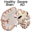
Alzheimer's disease brain severe.jpg 3,000 × 2,250; 3.86 MB
-
Alzheimers brain.jpg 500 × 492; 31 KB
-
Amygdala position.png 400 × 352; 69 KB
-
Anatomie-Basalganglien-A.jpg 2,026 × 1,010; 449 KB
-
Anatomie-Basalganglien.jpg 2,720 × 1,010; 458 KB
-
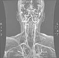
Angio MR.jpg 589 × 586; 58 KB
-
Arteriacerebral.jpg 200 × 176; 53 KB
-
ArteriaCerebral.jpg 274 × 212; 56 KB
-
Basal ganglia circuits.png 1,069 × 1,486; 709 KB
-
Basal ganglia in Parkinson's disease.png 1,061 × 1,046; 461 KB
-
Basal ganglia.jpg 564 × 507; 54 KB
-
Basal ganglia.png 3,452 × 1,500; 590 KB
-
Basal-ganglia-coronal-sections-large.png 800 × 350; 122 KB
-
Basal-ganglia-coronal-sections.png 487 × 217; 74 KB
-
Blausen 0104 Brain x-secs SectionalPlanes-ar.jpg 2,250 × 1,600; 463 KB
-
Blausen 0104 Brain x-secs SectionalPlanes-Arabic-YM.png 2,250 × 1,600; 2.21 MB
-
Blausen 0104 Brain x-secs SectionalPlanes.png 2,250 × 1,600; 1.85 MB
-
Bourgery & Jacob-cf16.jpg 1,785 × 2,574; 2.53 MB
-
Brain cut section 2.jpg 960 × 540; 83 KB
-
Brain herniation MRI.jpg 396 × 452; 29 KB
-
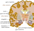
Brain human coronal section tags.png 628 × 413; 38 KB
-
Brain-41.jpg 248 × 250; 39 KB
-
Cingulate sulcus.png 170 × 140; 33 KB
-
Coronal cross-section of human brain.jpg 1,200 × 894; 158 KB
-
Coronal hippocampe.png 543 × 499; 298 KB
-
Coronal Image of a TOFI and a Normal Control.jpg 720 × 960; 70 KB
-
Coronal insula.png 560 × 409; 147 KB
-
Coupe d’un cerveau présentant une hémorragie cérébrale avec inondation ventriculaire.jpg 3,980 × 5,000; 5.21 MB
-
Cp coronale ssthal3.jpg 589 × 534; 74 KB
-
Cut section of human brain.jpg 960 × 540; 86 KB
-
Dichotisch.PNG 612 × 430; 39 KB
-
Dissociative identity disorder neuroscience brain imaging.png 696 × 466; 361 KB
-
Facial canal.png 480 × 538; 118 KB
-
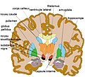
Frontal Section Basal Nuclei svg hariadhi.svg 512 × 512; 22 KB
-
Gehirn Frontalschnitt hippocampus-it.png 1,055 × 573; 247 KB
-
Gehirn Frontalschnitt hippocampus.png 913 × 573; 263 KB
-
Gray 718-amygdala.png 550 × 590; 310 KB
-
Gray 718-emphasizing-claustrum.png 550 × 590; 241 KB
-
Gray 718-emphasizing-corpus-callosum.png 550 × 590; 316 KB
-
Gray 718-emphasizing-putamen.png 550 × 590; 309 KB
-
Gray710.png 400 × 452; 46 KB
-
Gray717 without text.png 500 × 568; 92 KB
-
Gray717-emphasizing-insula.png 500 × 568; 294 KB
-
Gray717.png 500 × 568; 102 KB
-
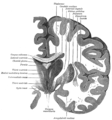
Gray718.png 550 × 590; 115 KB
-
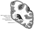
Gray738.png 450 × 376; 32 KB
-
Gray743.png 450 × 542; 45 KB
-
Gray744.png 550 × 481; 54 KB
-
Gray749.png 500 × 308; 22 KB
-
Gray759.png 354 × 550; 22 KB
-
Gray764 He.png 407 × 600; 75 KB
-
Gray764.png 407 × 600; 28 KB
-
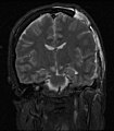
Head mri coronal section.jpg 577 × 925; 288 KB
-
Hemorragie intracerebrale.JPG 950 × 896; 389 KB
-
Hippocampe parahippo.png 1,000 × 800; 426 KB
-
Homunculus-ja.png 1,200 × 635; 197 KB
-
Homunculus.PNG 411 × 384; 112 KB
-
Hsv encephalitis.jpg 480 × 512; 88 KB
-
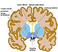
Human brain frontal (coronal) section description 2.JPG 702 × 487; 43 KB
-
Human brain frontal (coronal) section description.JPG 702 × 487; 43 KB
-
Human brain frontal (coronal) section description2.JPG 702 × 487; 42 KB
-
Human brain frontal (coronal) section.JPG 702 × 487; 41 KB
-
Human cerebral cortex.png 411 × 384; 84 KB
-
Human subventricular zone.jpg 1,200 × 553; 166 KB
-
Human temporal lobe areas.png 1,793 × 1,513; 1.63 MB
-
Huntington.jpg 722 × 902; 80 KB
-
MCA-Stroke-Brain-Human-1.JPG 720 × 540; 52 KB
-
MCA-Stroke-Brain-Human.JPG 720 × 540; 52 KB
-
Mesial Temporal Sclerosis.jpg 296 × 320; 24 KB
-
Motor homunculus-ja.png 558 × 561; 72 KB
-
Nervous and mental diseases (1908) (14591345130).jpg 1,828 × 1,312; 399 KB
-
PET2.jpg 360 × 359; 14 KB
-
Polymicrogyria arrows.JPG 610 × 598; 80 KB
-
PVNss.jpg 936 × 933; 133 KB
-
Sensory Homunculus.png 1,355 × 1,579; 55 KB
-
Slide10kk.JPG 960 × 720; 100 KB
-
Sobo 1909 642.png 1,063 × 1,019; 3.11 MB
-
Sobo 1909 645.png 1,227 × 750; 2.64 MB
-
Sobo 1909 646.png 1,201 × 773; 2.66 MB
-
Sobo 1909 681.png 1,005 × 877; 2.53 MB
-
Sobo 1911 643.png 2,492 × 1,544; 11.03 MB
-
Sobo 1911 644.png 2,496 × 1,692; 12.1 MB
-
Somatosensory cortex ja.png 1,200 × 1,240; 209 KB
-
SONss.jpg 903 × 944; 124 KB
-
T1map brain 3.png 256 × 256; 61 KB
-
TAC Brain tumor glioblastoma-Coronal plane.gif 512 × 512; 1.02 MB
-
The Journal of nervous and mental disease (1874) (14579735539).jpg 1,792 × 2,288; 403 KB
-
Tumor Germinoma Suprasellar3.JPG 575 × 728; 34 KB
-
U fibres big.JPG 1,638 × 1,458; 185 KB
-
VBM2.jpg 354 × 378; 16 KB
-
Ventral midbrain.png 994 × 1,017; 922 KB


