Category:Human axis
Jump to navigation
Jump to search
Subcategories
This category has the following 7 subcategories, out of 7 total.
3
- 3D data of human axis (2 F)
A
- Animations of human axis (5 F)
- Atlanto-axial joint (31 F)
O
- Ossification of human axis (3 F)
P
- Photographs of human axis (5 F)
Media in category "Human axis"
The following 66 files are in this category, out of 66 total.
-
De-Axis.ogg 1.6 s; 16 KB
-
 3D reconstructed CT image of C2 - a.png 920 × 754; 565 KB
-
 3D reconstructed CT image of C2 - b.png 1,078 × 954; 795 KB
-
 3D reconstructed CT image of C2 - c.png 1,145 × 845; 738 KB
-
 Atlasbogenspalt.jpg 1,142 × 1,031; 110 KB
-
 Axis (son).tif 991 × 503; 161 KB
-
 Axis Gr a.png 450 × 445; 34 KB
-
 Axis vertebra anterior view.jpg 956 × 876; 96 KB
-
 Axis vertebra posterior view.jpg 1,237 × 893; 138 KB
-
 Axis vertebrae.jpg 960 × 720; 79 KB
-
 BodyParts3D FJ6296 Axis.stl 5,120 × 2,880; 112 KB
-
 Braus 1921 70.png 1,344 × 1,227; 4.73 MB
-
 Braus 1921 71.png 732 × 411; 883 KB
-
 Braus 1921 72.png 1,179 × 678; 2.29 MB
-
 C1-C2 AP.JPG 2,303 × 1,800; 382 KB
-
 C1-C2 Lat.JPG 2,470 × 1,714; 302 KB
-
 C2 animation small.gif 320 × 320; 1.08 MB
-
 C2 back.png 900 × 900; 360 KB
-
 C2 from top animation small.gif 320 × 320; 1.4 MB
-
 C2 lateral.png 900 × 900; 332 KB
-
 Cervical vertebra 2 close-up back.png 900 × 900; 132 KB
-
 Cervical vertebra 2 close-up back2.png 900 × 900; 183 KB
-
 Cervical vertebra 2 close-up bottom animation.gif 600 × 600; 2.4 MB
-
 Cervical vertebra 2 close-up bottom.png 900 × 900; 166 KB
-
 Cervical vertebra 2 close-up frontal.png 900 × 900; 139 KB
-
 Cervical vertebra 2 close-up lateral animation.gif 600 × 600; 2 MB
-
 Cervical vertebra 2 close-up lateral.png 900 × 900; 122 KB
-
 Cervical vertebra 2 close-up top animation.gif 600 × 600; 2.21 MB
-
 Cervical vertebra 2 close-up top.png 900 × 900; 150 KB
-
 Condylar canal.jpg 708 × 518; 568 KB
-
 CT scan through a C2 specimen.png 1,862 × 696; 198 KB
-
 Cunningham’s Text-book of Anatomy (1914) - Fig 119.png 1,740 × 782; 850 KB
-
 Dens (odontoid process) of the axis.png 1,068 × 1,572; 844 KB
-
 Dens axis.jpg 864 × 970; 112 KB
-
 Denszielaufnahme mit geoeffnetem Mund 35M - CR - 001.jpg 1,319 × 1,106; 232 KB
-
 Dixon's Manual of human osteology (1912) - Fig 007.png 990 × 1,197; 688 KB
-
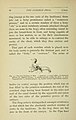
 DogAxisMalformation.jpg 1,197 × 817; 54 KB
-
 Gerrish's Text-book of Anatomy (1902) - Fig. 135.png 848 × 468; 237 KB
-
 Gerrish's Text-book of Anatomy (1902) - Fig. 136.png 1,049 × 456; 200 KB
-
 Gray105.png 416 × 164; 7 KB
-
 Gray305.png 575 × 504; 50 KB
-
 Gray306.png 450 × 250; 18 KB
-
 Gray307.png 600 × 416; 53 KB
-
 Gray308.png 500 × 485; 51 KB
-
 Gray387.png 443 × 600; 54 KB
-
 Gray86 zh.png 500 × 235; 45 KB
-
 Gray86-ar.png 910 × 428; 261 KB
-
 Gray86.png 500 × 235; 17 KB
-
 Gray87.heb.PNG 400 × 351; 19 KB
-
 Gray87.png 400 × 351; 21 KB
-
 Gray88.png 500 × 280; 15 KB
-
 Gray994 zh.png 600 × 861; 329 KB
-
 Gray994.png 600 × 861; 112 KB
-
 Human axis bone.stl 5,120 × 2,880; 11.04 MB
-
 Human Axis.stl 5,120 × 2,880; 4.65 MB
-
 HWS seitlich Annotation.jpg 754 × 1,100; 80 KB
-
 Ossification centers of the C2 vertebra.png 1,225 × 913; 303 KB
-
 Sobo 1909 186.png 1,192 × 992; 3.39 MB
-
 Sobo 1909 187.png 1,256 × 1,116; 4.02 MB
-
 Sobo 1909 188.png 1,344 × 1,048; 4.04 MB
-
 Sobo 1909 8.png 1,416 × 920; 3.73 MB
-
 The common frog (Page 68, Fig. 33) BHL7743449.jpg 2,099 × 3,308; 519 KB
-

-
 Vertebra - axis (lateral view).jpg 3,456 × 4,608; 3.03 MB
-
 Vertebra - axis (posterior).jpg 3,456 × 4,608; 3.64 MB
-
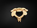 Vertebra - axis.jpg 4,608 × 3,456; 4.22 MB


_-_Fig_119.png)
_of_the_axis.png)


_-_Fig_007.png)

_-_Fig._135.png)
_-_Fig._136.png)


_BHL7743449.jpg)
_(14776278441).jpg)
.jpg)
.jpg)
