Category:Histopathology of breast ductal cell carcinomas in situ (DCIS)
Jump to navigation
Jump to search
Media in category "Histopathology of breast ductal cell carcinomas in situ (DCIS)"
The following 35 files are in this category, out of 35 total.
-
Breast DCIS Apocrine PA.JPG 3,070 × 2,301; 2.01 MB
-
Breast DCIS Comedonecrosis MP PA.JPG 3,070 × 2,809; 2.07 MB
-
Breast DCIS Comedonecrotic 2 PA.JPG 3,072 × 2,401; 1.04 MB
-
Breast DCIS Cribriform MP CTR.jpg 2,048 × 1,536; 792 KB
-
Breast DCIS Cribriform PA.JPG 3,070 × 2,531; 2.66 MB
-
Breast DCIS histopathology (1).jpg 500 × 376; 108 KB
-
Breast DCIS Micropapillary SNP.jpg 2,048 × 1,536; 815 KB
-
Breast DCIS MicropapillaryType MP CTR.jpg 2,048 × 1,536; 2.27 MB
-
Breast DCIS Mucinous Extravasation MP2 PA.JPG 2,597 × 2,745; 2.2 MB
-
Breast DCIS Mucinous Extravasation MP3 PA.JPG 2,875 × 2,809; 2.69 MB
-
Breast DCIS Papillary PA.JPG 3,070 × 2,301; 1.83 MB
-
Breast DCIS PapillaryVariant LP PA.JPG 2,367 × 2,301; 2.25 MB
-
Breast DCIS Solid IntermediateGrade SNP.jpg 2,048 × 1,536; 893 KB
-
Breast DCIS Solid PA.JPG 2,751 × 2,254; 1.75 MB
-
Breast DCIS Solid SNP.jpg 2,048 × 1,536; 1.47 MB
-
Breast Ductal Carcinoma in Situ With Calcifications (5436471236).jpg 1,449 × 1,448; 912 KB
-
Comedo DCIS.jpg 1,360 × 1,024; 816 KB
-
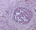
DCIS - Intraductal carcinoma of the breast.jpg 1,256 × 835; 511 KB
-
DCIS-2 (23266683042).jpg 1,234 × 839; 671 KB
-
DCIS-3 (23266684262).jpg 1,236 × 838; 700 KB
-
DCIS-4 (23079284350).jpg 1,236 × 840; 596 KB
-
DCIS-5 (22746705514).jpg 1,233 × 840; 419 KB
-
DCIS.jpg 647 × 542; 480 KB
-
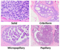
Histopathologic architectural patterns of DCIS.png 2,048 × 1,708; 5.68 MB
-
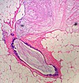
Histopathology of DCIS with lobular cancerization.jpg 1,119 × 1,193; 433 KB
-
Histopathology of ductal carcinoma in situ with comedo necrosis.jpg 2,048 × 1,532; 537 KB
-
Histopathology of high-grade DCIS, original.jpg 2,048 × 1,532; 444 KB
-
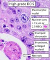
Histopathology of high-grade DCIS.png 1,415 × 1,677; 2.82 MB
-
Histopathology of microinvasive ductal carcinoma in situ, original.png 465 × 389; 497 KB
-
Histopathology of microinvasive ductal carcinoma in situ.png 466 × 387; 454 KB
-
Immunohistochemistry for E-cadherin in ductal carcinoma in situ.jpg 2,080 × 1,536; 789 KB
-
Immunohistochemistry for p120 in ductal carcinoma in situ.jpg 2,080 × 1,536; 893 KB
-
Immunohistochemistry for p120 in lobular carcinoma in situ.jpg 2,080 × 1,536; 783 KB
-
Intraductal Carcinoma.png 826 × 707; 1.17 MB
-
S10-5263 H&E 20x DCIS.jpg 1,360 × 1,024; 934 KB


