Category:Histopathology of breast cancer
Jump to navigation
Jump to search
Subcategories
This category has the following 6 subcategories, out of 6 total.
C
M
Media in category "Histopathology of breast cancer"
The following 44 files are in this category, out of 44 total.
-
Breast hyperplasia.jpg 960 × 717; 74 KB
-
Breast of rat with carcinogen ana alkaloids.jpg 960 × 717; 52 KB
-
Breast of rat with carcinogen.jpg 960 × 717; 58 KB
-
Collagenous spherulosis - high mag.jpg 4,272 × 2,848; 4.57 MB
-
Collagenous spherulosis - intermed mag.jpg 4,272 × 2,848; 4.85 MB
-
Collagenous spherulosis - very high mag.jpg 4,272 × 2,848; 4.23 MB
-
Cyclopædia of obstetrics and gynecology (1887) (14598138398).jpg 1,620 × 1,804; 939 KB
-
Cyclopædia of obstetrics and gynecology (1887) (14598265417).jpg 1,550 × 1,628; 760 KB
-
Cyclopædia of obstetrics and gynecology (1887) (14781631991).jpg 1,446 × 1,828; 450 KB
-
Cyclopædia of obstetrics and gynecology (1887) (14781645291).jpg 1,576 × 1,804; 576 KB
-
Cyclopædia of obstetrics and gynecology (1887) (14784431302).jpg 1,638 × 2,002; 915 KB
-
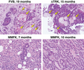
Figure 2 (6873190420).png 910 × 765; 1.4 MB
-
Figure 4B (7282699590).png 897 × 673; 1.54 MB
-
FISH Her2.jpg 1,280 × 1,280; 805 KB
-
HER2 FISH.jpg 1,600 × 838; 867 KB
-
Her2 staining on breast cancer tissue using antibody clone IHC002.jpg 1,200 × 900; 1.01 MB
-
Histopathology of basal-like breast cancer.jpg 3,079 × 2,047; 2.35 MB
-
Histopathology of secretory carcinoma, high magnification.jpg 1,481 × 1,532; 586 KB
-
Histopathology of secretory carcinoma, low magnification.jpg 2,048 × 1,532; 1.22 MB
-
IBC-D2-40.png 2,592 × 1,944; 8.01 MB
-
Lobular carcinoma in situ.jpg 1,360 × 1,024; 735 KB
-
Mammary myofibroblastoma - extra - intermed mag.jpg 4,272 × 2,848; 5.32 MB
-
Mammary myofibroblastoma - extra - very high mag.jpg 4,272 × 2,848; 7.08 MB
-
Mammary myofibroblastoma - high mag.jpg 4,272 × 2,848; 6.16 MB
-
Mammary myofibroblastoma - intermed mag.jpg 4,272 × 2,848; 5.46 MB
-
Mammary myofibroblastoma - low mag.jpg 4,272 × 2,848; 3.02 MB
-
Mammary myofibroblastoma - very high mag.jpg 4,272 × 2,848; 6.55 MB
-
Micrograph of ductal carcinoma with mild nuclear pleomorphism.jpg 552 × 507; 92 KB
-
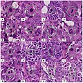
Mitosis appearances in breast cancer.jpg 1,595 × 1,594; 776 KB
-
Pie chart of histopathologic types of invasive breast cancer.png 2,058 × 1,195; 290 KB
-
Pie chart of histopathologic types of invasive breast cancer.svg 926 × 538; 18 KB
-
Pie chart of incidence and prognosis of histopathologic breast cancer types.png 2,795 × 1,220; 414 KB
-
Positive immunohistochemistry of KI-67 in invasive breast cancer.jpg 887 × 709; 200 KB
-
Radiographic Marker in Lumpectomy Specimen (3595823708).jpg 2,048 × 1,536; 994 KB
-
Radiographic Marker in Lumpectomy Specimen (3595823780).jpg 2,048 × 1,536; 1.08 MB
-
The breast- its anomalies, its diseases, and their treatment (1917) (14756710272).jpg 1,128 × 1,326; 708 KB
-
The breast- its anomalies, its diseases, and their treatment (1917) (14776918243).jpg 1,140 × 1,376; 703 KB
-
Tripolar Mitosis - breast carcinoma.jpg 2,048 × 1,536; 1.41 MB
-
Tubule formation score in the Nottingham system.jpg 602 × 201; 52 KB


