Category:Histology from CNX Anatomy & Physiology Textbook
Jump to navigation
Jump to search
Subcategories
This category has only the following subcategory.
Media in category "Histology from CNX Anatomy & Physiology Textbook"
The following 158 files are in this category, out of 158 total.
-
01 01ab Gross and Microscopic Anatomy.jpg 825 × 351; 243 KB
-
0319 Multinucleate Muscle Tissue Micrograph.jpg 710 × 487; 191 KB
-
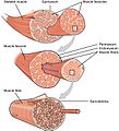
1002 Organization of Muscle Fiber.jpg 803 × 628; 363 KB
-
1007 Muscle Fibes (large) esp.jpg 740 × 770; 321 KB
-
1007 Muscle Fibes (large).jpg 675 × 741; 366 KB
-
1020 Cardiac Muscle gl.jpg 1,018 × 476; 121 KB
-
1020 Cardiac Muscle.jpg 1,018 × 476; 318 KB
-
1021 Smooth Muscle new.jpg 1,087 × 923; 416 KB
-
1022 Muscle Fibers (small) ku.jpg 819 × 610; 160 KB
-
1022 Muscle Fibers (small).jpg 801 × 642; 389 KB
-
1023 T-tubule (en).svg 624 × 372; 206 KB
-
1023 T-tubule (ru).svg 624 × 372; 207 KB
-
1023 T-tubule.jpg 577 × 368; 150 KB
-
1029 Smooth Muscle Motor Units.jpg 1,012 × 434; 236 KB
-
1316 Meningeal LayersN.jpg 949 × 515; 265 KB
-
1318b Dorsal Root Ganglion.jpg 1,621 × 921; 1.34 MB
-
1318b DRG.jpg 1,975 × 1,046; 1.44 MB
-
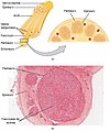
1319 Nerve Structure.jpg 902 × 1,060; 644 KB
-
1319 Nerve StructureN esp.jpg 895 × 1,069; 518 KB
-
1319 Nerve StructureN ukr.png 895 × 1,069; 1,002 KB
-
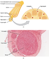
1319 Nerve StructureN.jpg 895 × 1,069; 677 KB
-
1319B Nerve Mag.jpg 1,975 × 958; 1.3 MB
-
1403 Olfaction-vi.jpg 2,196 × 2,258; 765 KB
-
1403 Olfaction.jpg 2,196 × 2,246; 1.58 MB
-
1403 Olfaction.png 2,059 × 2,106; 2.1 MB
-
1407 The Hair Cell.jpg 2,188 × 971; 803 KB
-
1414 Rods and Cones eng.jpg 1,050 × 1,542; 493 KB
-
1414 Rods and Cones.jpg 1,050 × 1,542; 646 KB
-
1427 Cochlea Micrograph.jpg 1,621 × 992; 868 KB
-
1504 Autonomic Varicosities.jpg 1,971 × 846; 549 KB
-
2004 Heart Wall.jpg 1,942 × 1,350; 530 KB
-
2017abc Cardiac Muscle-es.png 1,482 × 1,478; 1.11 MB
-
2017abc Cardiac Muscle.jpg 1,983 × 1,975; 1.29 MB
-
2102 Comparison of Artery and Vein.jpg 1,567 × 2,125; 2.88 MB
-
2103 Muscular and Elastic Artery Arteriole.jpg 2,271 × 579; 657 KB
-
2105 Capillary Bed esp.jpg 1,921 × 1,099; 422 KB
-
2105 Capillary Bed.jpg 1,859 × 1,099; 631 KB
-
2106 Large Medium Vein Venule pl.jpg 1,314 × 1,780; 528 KB
-
2106 Large Medium Vein Venule.jpg 1,314 × 1,780; 642 KB
-
2202 Lymphatic Capillaries big.png 587 × 362; 208 KB
-
2202 Lymphatic Capillaries small.png 521 × 499; 123 KB
-
2202 Lymphatic Capillaries.jpg 1,008 × 583; 331 KB
-
2204 The Hematopoietic System of the Bone Marrow new.jpg 2,251 × 1,828; 762 KB
-
2205 Bone Marrow.jpg 854 × 791; 505 KB
-
2206 The Location Structure and Histology of the Thymus.jpg 1,084 × 760; 502 KB
-
2207 Structure and Histology of a Lymph Node.jpg 1,093 × 407; 331 KB
-
2209 Location and Histology of Tonsils.jpg 910 × 1,173; 540 KB
-
2210 Mucosa Associated Lymphoid Tissue (MALT) Nodule.jpg 900 × 612; 416 KB
-
2230 Erythroblastosis Fetalis.jpg 1,105 × 1,009; 497 KB
-
2304 Pseudostratified Epithelium.jpg 1,931 × 1,131; 497 KB
-
2308 The Trachea-b.jpg 1,937 × 1,247; 544 KB
-
2309 The Respiratory Zone esp.jpg 2,188 × 1,671; 1.01 MB
-
2309 The Respiratory Zone.jpg 2,188 × 1,671; 1.26 MB
-
2310 Structures of the Respiratory Zone-b.jpg 1,759 × 1,039; 517 KB
-
2310 Structures of the Respiratory Zone.jpg 1,958 × 1,155; 1.02 MB
-
2311 Lung Tissue esp.jpg 1,737 × 903; 329 KB
-
2311 Lung Tissue.jpg 1,737 × 1,932; 1.02 MB
-
2402 Layers of the Gastrointestinal Tract.jpg 1,059 × 604; 336 KB
-
2409 Tooth.jpg 643 × 483; 203 KB
-
2415 Histology of StomachN esp.png 1,214 × 568; 477 KB
-
2415 Histology of StomachN.jpg 1,214 × 568; 415 KB
-
2418 Histology Small IntestinesN esp.jpg 2,158 × 1,648; 1.17 MB
-
2418 Histology Small IntestinesN.jpg 1,079 × 824; 540 KB
-
2421 Histology of the Large IntestineN.jpg 1,085 × 923; 608 KB
-
2424 Exocrine and Endocrine Pancreas FR.jpg 2,635 × 2,329; 643 KB
-
2424 Exocrine and Endocrine Pancreas-it.jpg 637 × 827; 367 KB
-
2424 Exocrine and Endocrine Pancreas.jpg 637 × 827; 441 KB
-
2605 The Bladder zh.jpg 1,962 × 1,202; 984 KB
-
2605 The Bladder.jpg 1,962 × 1,202; 1.09 MB
-
2607 Ureter.jpg 1,500 × 1,258; 2.25 MB
-
2611 Blood Flow in the Nephron.jpg 1,379 × 1,946; 1.06 MB
-
2612 Blood Flow in the Kidneys.jpg 1,971 × 1,565; 1.12 MB
-
2613 Podocytes.jpg 1,942 × 908; 884 KB
-
2614 Fenestrated Capillary esp.jpg 1,156 × 875; 293 KB
-
2614 Fenestrated Capillary.jpg 1,156 × 875; 351 KB
-
2615 Juxtaglomerular Apparatus.jpg 2,121 × 1,317; 1.27 MB
-
2616 Glomerulus.jpg 1,575 × 1,171; 992 KB
-
2901 Sperm Fertilization.jpg 1,910 × 1,318; 1.11 MB
-
401 Types of Tissue.jpg 1,070 × 1,135; 633 KB
-
403 Epithelial Tissue.jpg 1,209 × 970; 430 KB
-
404 Goblet Cell new-zh.jpg 2,539 × 4,722; 1.23 MB
-
404 Goblet Cell new.jpg 2,539 × 4,722; 1.26 MB
-
404b Goblet Cell new.jpg 2,299 × 2,231; 889 KB
-
405 Modes of Secretion by Glands Apocrine zh.png 1,342 × 543; 381 KB
-
405 Modes of Secretion by Glands Apocrine-es.png 1,400 × 567; 103 KB
-
405 Modes of Secretion by Glands Apocrine.png 1,342 × 543; 276 KB
-
405 Modes of Secretion by Glands updated.jpg 1,654 × 1,683; 816 KB
-
406 Types of Glands-es.png 1,164 × 1,260; 649 KB
-
406 Types of Glands.jpg 1,117 × 1,210; 568 KB
-
407 Sebaceous Glands esp.jpg 1,120 × 609; 383 KB
-
407 Sebaceous Glands.jpg 1,120 × 609; 464 KB
-
408 Connective Tissue-es.png 857 × 240; 309 KB
-
408 Connective Tissue.jpg 1,212 × 375; 358 KB
-
409 Adipose Tissue-es.png 2,044 × 820; 2.08 MB
-
409 Adipose Tissue.jpg 2,180 × 875; 1.1 MB
-
410 Reticular Tissue-es.png 1,868 × 699; 1.56 MB
-
410 Reticular Tissue.jpg 1,992 × 746; 1,003 KB
-
411 Reg Dense-Irregular Dense.jpg 1,972 × 1,372; 1.71 MB
-
412 Types of Cartilage-new.jpg 1,927 × 2,280; 2.14 MB
-
413 Types of Membranes.jpg 769 × 1,123; 355 KB
-
414 Skeletal Smooth Cardiac.jpg 635 × 1,068; 477 KB
-
414c Cardiacmuscle.jpg 591 × 403; 86 KB
-
415 Neuron.jpg 891 × 579; 303 KB
-
416 Nervous Tissue-new-es.png 2,246 × 847; 2.96 MB
-
416 Nervous Tissue-new.jpg 2,246 × 847; 1.87 MB
-
419 420 421 Table 04 01 updated.jpg 1,988 × 2,263; 1.91 MB
-
423 Table 04 02 Summary of Epithelial Tissue CellsN ku.jpg 558 × 895; 460 KB
-
423 Table 04 02 Summary of Epithelial Tissue CellsN.jpg 937 × 1,502; 564 KB
-
424 Blood A Fluid Connective Tissue-new.jpg 1,439 × 760; 443 KB
-
501 Structure of the skin.jpg 1,200 × 941; 652 KB
-
502 Layers of epidermis (no labels).png 792 × 647; 563 KB
-
502 Layers of epidermis.jpg 1,080 × 854; 656 KB
-
502ab Thin Skin versus Thick Skin.jpg 558 × 860; 352 KB
-
503 Epidermis.jpg 540 × 368; 207 KB
-
504 Melanocytes.jpg 1,133 × 894; 569 KB
-
505 Cells of the Epidermis.jpg 1,731 × 1,393; 1.01 MB
-
506 Hair.jpg 614 × 749; 256 KB
-
506 Layers of the Dermis.jpg 523 × 674; 494 KB
-
507 Nails.jpg 1,206 × 364; 211 KB
-
508 Eccrine gland.jpg 673 × 648; 318 KB
-
511 Hair Follicle.jpg 545 × 379; 317 KB
-
514 Light Micrograph of a Meissner Corpuscle.jpg 800 × 600; 476 KB
-
515 Thermoregulation.jpg 1,122 × 494; 463 KB
-
603 Anatomy of a Long Bone.jpg 684 × 1,156; 248 KB
-
603 Anatomy of Long Bone esp.jpg 800 × 1,200; 212 KB
-
603 Anatomy of Long Bone zh.jpg 708 × 1,156; 152 KB
-
603 Anatomy of Long Bone.jpg 708 × 1,156; 257 KB
-
604 Bone cells esp.jpg 807 × 600; 155 KB
-
604 Bone cells.jpg 807 × 567; 198 KB
-
605 Compact Bone esp.jpg 1,200 × 850; 428 KB
-
605 Compact Bone.jpg 1,181 × 798; 477 KB
-
606 Spongy Bone esp.jpg 1,119 × 743; 289 KB
-
606 Spongy Bone.jpg 1,119 × 743; 359 KB
-
607 Periosteum and Endosteum.jpg 1,071 × 535; 254 KB
-
608 Endochrondal Ossification.jpg 1,133 × 1,512; 487 KB
-
609 Body Supply to the Bone.jpg 582 × 810; 220 KB
-
611 Intramembraneous Ossification.jpg 1,213 × 847; 672 KB
-
619 Red and Yellow Bone Marrow.jpg 679 × 418; 214 KB
-
621 Anatomy of a Flat Bone esp.jpg 812 × 411; 104 KB
-
621 Anatomy of a Flat Bone.jpg 812 × 411; 135 KB
-
622 Longitudinal Bone Growth.jpg 800 × 1,246; 493 KB
-
623 Epiphyseal Plate-Line.jpg 790 × 625; 157 KB
-
624 Diagram of Compact Bone-new.jpg 1,413 × 1,664; 1.05 MB
-
906 Cartiliginous Joints.jpg 2,274 × 1,078; 706 KB
-
907 Synovial Joints (ro).jpg 1,425 × 1,578; 408 KB
-
907 Synovial Joints.jpg 1,425 × 1,578; 725 KB
-
Aufbau Skelettmuskulatur deutsch.png 704 × 741; 406 KB
-
Bindegewebe.png 593 × 373; 229 KB
-
Juxtaglomerular Apparatus and Glomerulus esp.jpg 1,954 × 724; 615 KB
-
Juxtaglomerular Apparatus and Glomerulus numbers.png 2,257 × 897; 1.61 MB
-
Juxtaglomerular Apparatus and Glomerulus pl.png 2,443 × 905; 1.7 MB
-
Juxtaglomerular Apparatus and Glomerulus-zh.jpg 1,954 × 724; 392 KB
-
Juxtaglomerular Apparatus and Glomerulus.jpg 1,954 × 724; 801 KB
-
Layers of the GI Tract english.svg 700 × 431; 516 KB
-
Layers of the GI Tract es.svg 680 × 443; 848 KB
-
Layers of the GI Tract numbers.svg 587 × 452; 505 KB
-
Meningeal Layers fa.jpg 800 × 434; 93 KB
-
Simple columnar epithelial cells.png 244 × 132; 31 KB


