Category:Histological schematic
Jump to navigation
Jump to search
Subcategories
This category has the following 8 subcategories, out of 8 total.
B
D
E
R
S
Media in category "Histological schematic"
The following 100 files are in this category, out of 100 total.
-
406 Types of Glands-es.png 1,164 × 1,260; 649 KB
-
406 Types of Glands.jpg 1,117 × 1,210; 568 KB
-
408 Connective Tissue-es.png 857 × 240; 309 KB
-
408 Connective Tissue.jpg 1,212 × 375; 358 KB
-
409 Adipose Tissue-es.png 2,044 × 820; 2.08 MB
-
409 Adipose Tissue.jpg 2,180 × 875; 1.1 MB
-
410 Reticular Tissue-es.png 1,868 × 699; 1.56 MB
-
410 Reticular Tissue.jpg 1,992 × 746; 1,003 KB
-
412 Types of Cartilage-new.jpg 1,927 × 2,280; 2.14 MB
-
416 Nervous Tissue-new-es.png 2,246 × 847; 2.96 MB
-
416 Nervous Tissue-new.jpg 2,246 × 847; 1.87 MB
-
419 420 421 Table 04 01 updated.jpg 1,988 × 2,263; 1.91 MB
-
Anatomical pathology.svg 1,888 × 1,038; 120 KB
-
Bindegewebe.png 593 × 373; 229 KB
-
Chem. Synapse scheme.jpg 200 × 212; 12 KB
-
Cortis organ stor.png 800 × 316; 47 KB
-
Detail de coupe longitudinale de tête d'embryon de souris.jpg 3,120 × 4,160; 3.09 MB
-
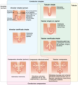
Epitely.svg 1,123 × 794; 116 KB
-
Esquema discos intercalares.jpg 1,736 × 1,089; 198 KB
-
Extracellular Matrix numbers.png 1,122 × 603; 86 KB
-
Extracellular Matrix-es.png 1,183 × 792; 69 KB
-
Extracellular Matrix.png 1,262 × 845; 25 KB
-
Gonadenanlage1.svg 720 × 1,920; 187 KB
-
Gray1063.png 600 × 350; 51 KB
-
Gray1064.png 400 × 474; 49 KB
-
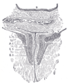
Gray1071.png 350 × 409; 29 KB
-
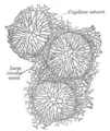
Gray1072.png 200 × 237; 7 KB
-
Gray1074.png 500 × 363; 79 KB
-
Gray1079.png 400 × 479; 62 KB
-
Gray1082-ar.png 500 × 367; 93 KB
-
Gray1082.png 500 × 367; 62 KB
-
Gray1089.png 400 × 495; 60 KB
-
Gray1090.png 400 × 448; 56 KB
-
Gray1093-ar.png 400 × 403; 126 KB
-
Gray1093.png 400 × 403; 35 KB
-
Gray1094.png 300 × 256; 14 KB
-
Gray11.png 500 × 178; 12 KB
-
Gray1105-ar.png 426 × 400; 167 KB
-
Gray1105.png 426 × 400; 42 KB
-
Gray1106.png 1,090 × 849; 510 KB
-
Gray1113 zh.png 499 × 373; 40 KB
-
Gray1113-ar.png 600 × 373; 77 KB
-
Gray1113.png 600 × 373; 40 KB
-
Gray1114.png 430 × 450; 44 KB
-
Gray1126.png 460 × 500; 34 KB
-
Gray1128.png 602 × 600; 43 KB
-
Gray1132.png 500 × 400; 42 KB
-
Gray1133.png 453 × 300; 44 KB
-
Gray1141-ar.png 366 × 500; 190 KB
-
Gray1141.png 366 × 500; 49 KB
-
Gray1145.png 661 × 500; 64 KB
-
Gray1150 zh.png 483 × 250; 59 KB
-
Gray1150.png 843 × 485; 198 KB
-
Gray1157.png 433 × 400; 35 KB
-
Gray1191 zh.png 401 × 300; 80 KB
-
Gray1191.png 401 × 300; 34 KB
-
Gray1192.png 578 × 300; 27 KB
-
Gray15.png 400 × 485; 79 KB
-
Gray33.png 298 × 625; 28 KB
-
Gray374.png 172 × 300; 13 KB
-
Gray39 cs.png 576 × 384; 50 KB
-
Gray39 sp.PNG 500 × 384; 38 KB
-
Gray39.png 500 × 384; 41 KB
-
Gray706-vi.png 622 × 638; 162 KB
-
Gray706.png 550 × 598; 56 KB
-
Gray754.png 500 × 649; 65 KB
-
Gray81.png 407 × 250; 18 KB
-
Gray893 - sweat glands.png 253 × 500; 49 KB
-
Gray964.png 402 × 600; 78 KB
-
Histological section-it.svg 591 × 781; 157 KB
-
Histologie der Nebenniere.png 477 × 454; 42 KB
-
HumanTissues.jpg 960 × 720; 73 KB
-
Hyperplasia vs Hypertrophy pl.png 1,471 × 1,776; 501 KB
-
Hyperplasia vs Hypertrophy-tr.png 706 × 852; 340 KB
-
Hyperplasia vs Hypertrophy.svg 706 × 852; 206 KB
-
Hypertrophy.jpg 512 × 384; 15 KB
-
Illu lymph node structure az.jpg 520 × 300; 35 KB
-
Illu lymph node structure de.png 520 × 300; 97 KB
-
Illu vein.jpg 352 × 150; 21 KB
-
Matriz extracelular.png 1,245 × 725; 141 KB
-
Membrane basale.jpg 472 × 383; 27 KB
-
Meyers b11 s0063.jpg 800 × 1,275; 425 KB
-
Meyers b7 s0237.jpg 800 × 1,275; 671 KB
-
MucoseCellNumbered.svg 400 × 400; 28 KB
-
Nenadorove zmeny.svg 720 × 405; 34 KB
-
Neuron labels ca.png 400 × 215; 40 KB
-
Neuron.jpg 460 × 314; 42 KB
-
Non-neoplastic changes.svg 720 × 405; 34 KB
-
NonneoplasidrawDH.JPG 653 × 257; 22 KB
-
Piel y cabello vs feolamina.jpg 300 × 275; 27 KB
-
Plazenta.png 500 × 384; 40 KB
-
Pseudostratified Columnar Epithelium – Numbered.svg 720 × 720; 48 KB
-
Přehledné barvicí metody.svg 1,123 × 794; 67 KB
-
Schema Alveole.jpg 600 × 486; 102 KB
-
Schéma erytrocytu.svg 294 × 132; 10 KB
-
SerouseCellNumbered.svg 400 × 400; 36 KB
-
Siebert 11.jpg 1,934 × 2,858; 1.3 MB
-
The wall of the large intestine (diagram).jpg 595 × 842; 100 KB
-
Transverseureter.png 464 × 500; 57 KB
-
Zab.jpg 539 × 652; 219 KB
