Category:Functional magnetic resonance imaging
Jump to navigation
Jump to search
Subcategories
This category has only the following subcategory.
Media in category "Functional magnetic resonance imaging"
The following 85 files are in this category, out of 85 total.
-
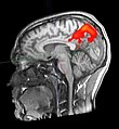
Aktivitaethinten.jpg 220 × 239; 24 KB
-
BOLD-Granger-Causality-Reflects-Vascular-Anatomy-pone.0084279.s001.ogv 2.6 s, 510 × 480; 461 KB
-
Brain network dynamics in creativity.webp 2,100 × 610; 94 KB
-
-
-
CFS-brain-scan-basal-ganglia-fMRI.png 1,483 × 921; 714 KB
-
CO2-O2-fMRI-all-over-time.png 2,800 × 4,269; 5.34 MB
-
-
-
-
-
-
-
-
-
Effects-of-thresholding-on-correlation-based-image-similarity-metrics-Video1.ogv 2.0 s, 3,200 × 1,537; 328 KB
-
Effects-of-thresholding-on-correlation-based-image-similarity-metrics-Video2.ogv 2.0 s, 3,200 × 1,537; 317 KB
-
FEAT.png 435 × 446; 30 KB
-
Fingertapping experiment DXIII.jpg 640 × 640; 110 KB
-
FMRI BOLD activation in an emotional Stroop task.jpg 886 × 655; 366 KB
-
FMRI coronal scan of Nathanial Bradford.png 709 × 744; 1.08 MB
-
FMRI of Nathanial Bradford.png 709 × 637; 814 KB
-
FMRI V1 RSFC LSD.png 495 × 671; 342 KB
-
FMRI-scan sectie 85.JPG 512 × 510; 59 KB
-
FMRIScan.jpg 480 × 418; 118 KB
-
Fnbeh-08-00171-g002.jpg 850 × 636; 191 KB
-
FSL overview.png 719 × 625; 102 KB
-
FSL spatialsmoothing screenshot1.png 586 × 478; 83 KB
-
FSL start GUI and motion correction buttion.png 663 × 491; 83 KB
-
FSL toolbar.png 538 × 91; 20 KB
-
Functional magnetic resonance imaging.jpg 250 × 208; 11 KB
-
Functional-MRI-in-Awake-Unrestrained-Dogs-pone.0038027.s006.ogv 5 min 44 s, 853 × 480; 48.13 MB
-
Fusiform face area face recognition.jpg 475 × 503; 95 KB
-
Física y fisiología de la señal BOLD de fMRI.svg 850 × 500; 48 KB
-
Heine2012x3010.png 3,010 × 2,920; 4.05 MB
-
High Resolution FMRI of the Human Brain.gif 512 × 256; 1.06 MB
-
Human-visual-cortical-responses-to-specular-and-matte-motion-flows-Video1.ogv 15 s, 512 × 512; 1.2 MB
-
Human-visual-cortical-responses-to-specular-and-matte-motion-flows-Video2.ogv 15 s, 512 × 512; 1.38 MB
-
Human-visual-cortical-responses-to-specular-and-matte-motion-flows-Video3.ogv 15 s, 512 × 512; 1.5 MB
-
Human-visual-cortical-responses-to-specular-and-matte-motion-flows-Video4.ogv 15 s, 512 × 512; 2.65 MB
-
Human-visual-cortical-responses-to-specular-and-matte-motion-flows-Video5.ogv 2.1 s, 512 × 512; 224 KB
-
Human-visual-cortical-responses-to-specular-and-matte-motion-flows-Video6.ogv 4.1 s, 512 × 512; 81 KB
-
Human-visual-cortical-responses-to-specular-and-matte-motion-flows-Video7.ogv 4.1 s, 512 × 512; 147 KB
-
Human-visual-cortical-responses-to-specular-and-matte-motion-flows-Video8.ogv 10 s, 1,440 × 1,200; 401 KB
-
Human-visual-cortical-responses-to-specular-and-matte-motion-flows-Video9.ogv 10 s, 1,440 × 1,200; 3.78 MB
-
MI fMRI.jpg 517 × 387; 96 KB
-
MVPA and fMRI-Adaptation.png 1,386 × 532; 514 KB
-
NPH MRI 266 GILD.gif 256 × 256; 244 KB
-
NPH MRI 267 GILD.gif 256 × 256; 437 KB
-
NPH MRI 268 GILD.gif 256 × 256; 585 KB
-
NPH MRI 272 GILD.gif 256 × 256; 639 KB
-
Powell2004Fig1A.jpeg 392 × 486; 35 KB
-
Powell2004Fig1A.png 326 × 206; 81 KB
-
Rapid-Amygdala-Responses-during-Trace-Fear-Conditioning-without-Awareness-pone.0096803.s002.ogv 25 s, 1,440 × 1,080; 3.07 MB
-
Real-Time-fMRI-Pattern-Decoding-and-Neurofeedback-Using-FRIEND-An-FSL-Integrated-BCI-Toolbox-pone.0081658.s001.ogv 1 min 24 s, 1,440 × 1,080; 10.48 MB
-
Real-Time-fMRI-Pattern-Decoding-and-Neurofeedback-Using-FRIEND-An-FSL-Integrated-BCI-Toolbox-pone.0081658.s002.ogv 56 s, 1,440 × 1,080; 15.93 MB
-
Real-Time-fMRI-Pattern-Decoding-and-Neurofeedback-Using-FRIEND-An-FSL-Integrated-BCI-Toolbox-pone.0081658.s003.ogv 1 min 31 s, 1,280 × 720; 9.23 MB
-
Repetition Suppression (RS) Models.png 1,078 × 720; 418 KB
-
Resonancia Funcional.jpg 297 × 170; 10 KB
-
Resonancia magnética Funcional - 1.wav 52 s; 8.81 MB
-
Resonancia magnética Funcional - 2.wav 52 s; 8.78 MB
-
Resting-state functional connectivity effects of Floatation-REST.jpg 669 × 413; 56 KB
-
Retinotopic English.jpg 1,476 × 945; 427 KB
-
Schematic of cortical areas involved with pain processing and fMRI cropped.jpg 1,172 × 792; 211 KB
-
Schematic of cortical areas involved with pain processing and fMRI.jpg 1,200 × 1,383; 378 KB
-
Temporal-Non-Local-Means-Filtering-Reveals-Real-Time-Whole-Brain-Cortical-Interactions-in-Resting-pone.0158504.s002.ogv 2 min 20 s, 330 × 240; 10.66 MB
-
Temporal-Non-Local-Means-Filtering-Reveals-Real-Time-Whole-Brain-Cortical-Interactions-in-Resting-pone.0158504.s003.ogv 2 min 20 s, 330 × 240; 16.92 MB
-
Temporal-Non-Local-Means-Filtering-Reveals-Real-Time-Whole-Brain-Cortical-Interactions-in-Resting-pone.0158504.s004.ogv 2 min 20 s, 330 × 240; 8.15 MB
-
-
-
-
-
-
Ventral midbrain.png 994 × 1,017; 922 KB


