Category:Files from Ed Uthman Flickr stream uploaded by Netha Hussain
Jump to navigation
Jump to search
Media in category "Files from Ed Uthman Flickr stream uploaded by Netha Hussain"
The following 200 files are in this category, out of 385 total.
(previous page) (next page)-
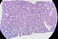
 23-year-old woman, routine Pap (4055895532).jpg 1,338 × 892; 671 KB
-
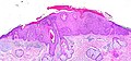 Actinic Keratosis (2230218979).jpg 1,733 × 804; 751 KB
-
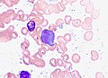
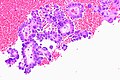
 Acute Promyelocytic Leukemia, Marrow Aspirate (40160031375).jpg 1,974 × 1,186; 573 KB
-
 Acute Promyelocytic Leukemia, Marrow Aspirate (41052768711).jpg 1,164 × 927; 273 KB
-
 Acute Promyelocytic Leukemia, Marrow Aspirate (41052769041).jpg 1,344 × 977; 315 KB
-
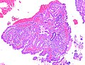
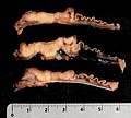
 Adenocarcinoma of the Cecum (52241313371).jpg 2,289 × 4,004; 4.51 MB
-
 Adenocarcinoma of the Sigmoid Colon (51315163832).jpg 3,305 × 2,759; 3.17 MB
-
 Adenomyosis, Hysterectomy Specimen (640264025).jpg 4,229 × 2,518; 3.01 MB
-
 Amyloidosis, H&E (377490167).jpg 876 × 714; 368 KB
-
 Amyloidosis, H&E (377490242).jpg 905 × 762; 435 KB
-
 Amyloidosis, H&E (377490371).jpg 876 × 723; 426 KB
-
 Amyloidosis, Inguinal Node, Gross (377533391).jpg 3,522 × 2,830; 3.47 MB
-
 Amyloidosis, Node, Congo Red (377559857).jpg 818 × 743; 267 KB
-
 Arias-Stella Reaction in Endometrium (45966643982).jpg 2,301 × 2,017; 2.18 MB
-
 Arterial Thrombus (51955463785).jpg 1,767 × 1,981; 1.71 MB
-
 Axillary Lymph Node with Black (Tattoo?) Pigment (17509353414).jpg 2,048 × 1,536; 1,013 KB
-
 Basal Cell Adenoma of the Parotid Gland (51101942534).jpg 1,658 × 2,155; 1.98 MB
-
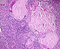
 Basal Cell Adenoma, Parotid gland (34490880674).jpg 1,275 × 943; 327 KB
-
 Basal Cell Adenoma, Parotid gland (34491007474).jpg 1,998 × 1,700; 1.23 MB
-
 Basal Cell Adenoma, Parotid gland (34946909600).jpg 2,528 × 1,616; 1.66 MB
-
 Basal Cell Adenoma, Parotid gland (35167484102).jpg 2,563 × 1,608; 1.57 MB
-
 Basal Cell Adenoma, Parotid gland (35167486672).jpg 2,563 × 2,126; 1.86 MB
-
 Basal Cell Adenoma, Parotid gland (35204667831).jpg 2,550 × 1,029; 1,005 KB
-
 Basal Cell Adenoma, Parotid gland (35204669261).jpg 2,302 × 1,264; 1.13 MB
-
 Basal Cell Adenoma, Parotid gland (35204673571).jpg 2,515 × 1,703; 1.75 MB
-
 Basal Cell Adenoma, Parotid gland (35204774571).jpg 2,346 × 1,638; 1.27 MB
-
 Basal Cell Adenoma, Parotid gland (35334422745).jpg 2,555 × 2,130; 1.62 MB
-
 Basal Cell Adenoma, Parotid gland (35334423775).jpg 2,620 × 2,035; 1.9 MB
-
 Bilobate Placenta with Velamentous Insertion of the Cord (8271783612).jpg 1,936 × 2,592; 1.31 MB
-
 Brisk Extravascular Hemolytic Anemia (24467236849).jpg 1,428 × 1,426; 1 MB
-
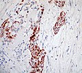 Calretinin (+) (2164275352).jpg 849 × 754; 365 KB
-
 CAM 5.2 (+) (2164276824).jpg 1,024 × 768; 272 KB
-
 Carcinoma of the Breast Invading Pacinian Corpuscle in Overlying Skin (51503787366).jpg 2,018 × 2,282; 2.73 MB
-
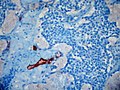 CD10 (-) (2163477587).jpg 1,024 × 768; 483 KB
-
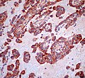 CD99 (+) (2164277470).jpg 821 × 754; 366 KB
-
 Cervical biopsy, 200X (4055154081).jpg 1,677 × 1,119; 627 KB
-
 Cervical biopsy, 400X (4055154115).jpg 1,735 × 1,158; 724 KB
-
 Cervical biopsy, Ki-67 immunostain (4055154159).jpg 1,605 × 1,072; 1,011 KB
-
 Charcot-Leyden Crystals (51787695298).jpg 1,910 × 1,494; 3.17 MB
-
 Cholesteatoma, Gross Specimen (51144665835).jpg 2,317 × 2,065; 1.24 MB
-
 Cholesterol Granuloma of the Nasal Cavity (27085223157).jpg 2,100 × 1,515; 1.59 MB
-
 Cholesterol Granuloma of the Nasal Cavity (27085225477).jpg 2,154 × 2,071; 1.86 MB
-
 Cholesterol Granuloma of the Nasal Cavity (27085226697).jpg 1,990 × 1,905; 2 MB
-
 Cholesterol Granuloma of the Nasal Cavity (40147206780).jpg 2,526 × 2,512; 3.22 MB
-
 Cholesterol Granuloma of the Nasal Cavity (41953897841).jpg 2,215 × 1,992; 1.94 MB
-
 Cholesterol polyp in the gallbladder (50227720502).jpg 1,979 × 2,336; 2.58 MB
-
 Chronic Myelomonocytic Leukemia, Peripheral Blood Smear (50506617237).jpg 2,340 × 1,880; 6.01 MB
-
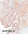
 Clear Cell Carcinoma of the Endometrium (32860175497).jpg 1,787 × 2,403; 3.08 MB
-
 Clear Cell Carcinoma of the Endometrium (33926551598).jpg 1,850 × 1,989; 1.55 MB
-
 Clear Cell Carcinoma of the Endometrium (33926629198).jpg 1,742 × 1,879; 1.82 MB
-
 Clear Cell Carcinoma of the Endometrium (33926684288).jpg 1,336 × 1,947; 1.56 MB
-
 Clear Cell Carcinoma of the Endometrium (46887120155).jpg 1,801 × 2,035; 1.59 MB
-
 Clear Cell Carcinoma of the Endometrium (46887232725).jpg 1,852 × 1,368; 1.25 MB
-
 Clear Cell Carcinoma of the Endometrium (46887233135).jpg 1,962 × 1,500; 1.35 MB
-
 Clear Cell Carcinoma of the Endometrium (46887233345).jpg 1,556 × 1,551; 1.36 MB
-
 Clear Cell Carcinoma of the Endometrium (47014344944).jpg 1,223 × 2,296; 1.74 MB
-
 Clear Cell Carcinoma of the Endometrium (47751406072).jpg 1,575 × 1,930; 1.26 MB
-
 Clear Cell Carcinoma of the Endometrium (47751407452).jpg 1,707 × 2,078; 1.78 MB
-
 Clear Cell Carcinoma of the Endometrium (47751407572).jpg 1,922 × 1,935; 1.76 MB
-
 Clear Cell Carcinoma of the Endometrium (47751482882).jpg 1,667 × 1,116; 747 KB
-
 Clear Cell Carcinoma of the Endometrium (47751540062).jpg 1,814 × 1,535; 1.62 MB
-
 Clear Cell Carcinoma of the Endometrium (47803494281).jpg 1,446 × 2,107; 1.26 MB
-
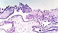
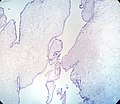
 Collagenous Gastritis (43856191530).jpg 2,670 × 1,376; 2.17 MB
-
 Collagenous Gastritis (44949450304).jpg 2,573 × 1,472; 1.81 MB
-
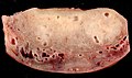
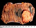
 Colon Adenocarcinoma (gross) NZ7 1386 (51440373932).jpg 6,347 × 4,328; 24.28 MB
-
 Colon Adenocarcinoma (gross) NZ7 1388 (51441869729).jpg 6,119 × 4,826; 28.42 MB
-
 Colon Adenocarcinoma (gross) NZ7 1389 (51441868574).jpg 3,770 × 3,379; 9.12 MB
-
 Colon Adenocarcinoma 2 (225253293).jpg 4,288 × 2,848; 6.81 MB
-
 Colon Adenocarcinoma 3 (225253295).jpg 3,868 × 2,655; 3.94 MB
-
 Colon Adenocarcinoma 4 (225253297).jpg 4,288 × 2,848; 5.08 MB
-
 Complete Hydatidiform Mole (25650505087).jpg 2,740 × 1,646; 1.53 MB
-
 Complete Hydatidiform Mole (25651404517).jpg 2,155 × 1,243; 892 KB
-
 Complete Hydatidiform Mole (25651405367).jpg 1,958 × 1,730; 972 KB
-
 Complete Hydatidiform Mole (26636232238).jpg 6,048 × 4,032; 12.21 MB
-
 Complete Hydatidiform Mole (26649804828).jpg 2,967 × 2,448; 2.57 MB
-
 Complete Hydatidiform Mole (26649819168).jpg 2,368 × 2,295; 1.61 MB
-
 Complete Hydatidiform Mole (26650631648).jpg 1,841 × 1,530; 794 KB
-
 Complete Hydatidiform Mole (38711522330).jpg 2,598 × 2,426; 1.77 MB
-
 Complete Hydatidiform Mole (38711522360).jpg 2,581 × 2,396; 1.87 MB
-
 Complete Hydatidiform Mole (38711524440).jpg 2,015 × 1,748; 1.05 MB
-
 Complete Hydatidiform Mole (38711526030).jpg 1,732 × 2,048; 1.34 MB
-
 Complete Hydatidiform Mole (39611782015).jpg 6,048 × 4,032; 12.08 MB
-
 Complete Hydatidiform Mole (39611785555).jpg 4,259 × 3,948; 7.47 MB
-
 Complete Hydatidiform Mole (39611790275).jpg 6,048 × 4,032; 11.79 MB
-
 Complete Hydatidiform Mole (39626392205).jpg 2,063 × 1,491; 872 KB
-
 Complete Hydatidiform Mole (39810634804).jpg 1,862 × 2,165; 1.48 MB
-
 Complete Hydatidiform Mole (40478281822).jpg 3,264 × 2,448; 2.82 MB
-
 Complete Hydatidiform Mole (40479191442).jpg 2,033 × 1,921; 986 KB
-
 Complete Hydatidiform Mole (40479194942).jpg 2,524 × 2,448; 1.6 MB
-
 Complete Hydatidiform Mole (51479531243).jpg 816 × 960; 100 KB
-
 Conjunctival Melanoma (1857361150).jpg 2,131 × 1,421; 1.61 MB
-
 Cryptococcus in cerebrospinal fluid, India ink prep. (49623531757).jpg 1,659 × 1,938; 1.26 MB
-
 Cryptococcus Yeasts in Circulating Granulocytes (51754021280).jpg 813 × 806; 79 KB
-
 Cutaneous Keratocyst (50267985312).jpg 2,107 × 2,124; 1.99 MB
-
 Cystic Lymphangioma of the Cecum (42737223084).jpg 2,894 × 2,467; 1.71 MB
-
 Cystic Pilomatricoma (52304203643).jpg 1,785 × 1,357; 775 KB
-
 Cytokeratins AE1-AE3 (+) (2163477507).jpg 791 × 754; 329 KB
-
 Desmin (+) (2163477293).jpg 1,024 × 768; 312 KB
-
 Digital Mucous Cyst (24741722691).jpg 2,048 × 1,536; 644 KB
-
 Digital Mucous Cyst, Foot (20127190050).jpg 1,693 × 1,127; 540 KB
-
 Digital Mucous Cyst, Toe (19338000950).jpg 1,504 × 1,426; 672 KB
-
 DSCN3258 (22334543716).jpg 2,048 × 1,536; 768 KB
-
 DSCN3260 (22371250631).jpg 2,048 × 1,536; 710 KB
-
 DSCN3264 (22173703859).jpg 2,048 × 1,536; 714 KB
-
 DSCN3271 (22371246701).jpg 2,048 × 1,536; 657 KB
-
 DSCN3281 smooth muscle actin (SMA) (21739375573).jpg 1,689 × 1,124; 913 KB
-
 DSCN3282 DOG-1 (22360485835).jpg 1,687 × 1,124; 829 KB
-
 E.T. The Umbilical Cord (3276268079).jpg 1,828 × 1,498; 1.48 MB
-
 Embolized Uterus (44319080955).jpg 1,270 × 1,124; 808 KB
-
 Embolized Uterus (44319081065).jpg 1,352 × 1,124; 811 KB
-
 Embolized Uterus (44507244114).jpg 1,350 × 1,124; 646 KB
-
 Embolized Uterus (44507244194).jpg 1,124 × 1,208; 885 KB
-
 Empty Gestational Sac in a Tubal Pregnancy (27310950457).jpg 2,164 × 1,956; 1.89 MB
-
 Endolymphatic Sac Tumor 1 (231907666).jpg 1,280 × 960; 977 KB
-
 Endolymphatic Sac Tumor 2 (231907667).jpg 1,280 × 960; 1.03 MB
-
 Endolymphatic Sac Tumor 3 (231907669).jpg 1,280 × 960; 1,018 KB
-
 Endometrial Intraepithelial Neoplasia (EIN) (46262991251).jpg 2,347 × 1,706; 1.61 MB
-
 Eosinophils in Peripheral Blood Smear (4746281402).jpg 1,426 × 1,266; 1.38 MB
-
 Estrogen receptor (+) (2164277240).jpg 1,024 × 768; 703 KB
-
 Food particle from a colonoscopy specimen (34016474661).jpg 1,480 × 811; 369 KB
-
 Foreign Body Granuloma on the Peritoneum (41962302110).jpg 2,402 × 1,458; 1.28 MB
-
 Foreign Material in Bone Marrow (H&E, 1000X, oil) (4837907128).jpg 1,587 × 1,309; 768 KB
-
 Foreign Material in Bone Marrow (H&E, 100X) (4837290693).jpg 1,239 × 1,127; 572 KB
-
 Foreign Material in Bone Marrow (H&E, 200X) (4837292483).jpg 1,555 × 1,353; 755 KB
-
 Foreign Material in Bone Marrow (H&E, 400X) (4837293823).jpg 1,443 × 1,386; 838 KB
-
 Foreign Material in Bone Marrow (H&E, crossed polarizers, 400X) (4837906214).jpg 1,476 × 1,254; 836 KB
-
 Fundic Gland Polyp of Stomach (20704194942).jpg 1,210 × 806; 405 KB
-
 Fundic Gland Polyp of the Stomach (49250095622).jpg 2,108 × 1,397; 1.29 MB
-
 Fungus Ball (51003223298).jpg 2,114 × 2,178; 4.08 MB
-
 Fungus Ball (51003927106).jpg 2,133 × 1,938; 2.51 MB
-
 Fungus Ball, Sphenoid Sinus (51749745479).jpg 1,864 × 2,186; 2.04 MB
-
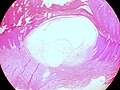
 Ganglion Cyst (27091675117).jpg 2,463 × 1,558; 1.16 MB
-
 Ganglion Cyst of Hand (35295313226).jpg 2,102 × 1,669; 659 KB
-
 GIST Removed from the stomach, gross external appearance (21959091348).jpg 1,865 × 1,840; 1.39 MB
-
 Glomangioma (50227721402).jpg 1,967 × 2,207; 3.26 MB
-
 Glomangioma (Glomuvenous Malformation) (48879295668).jpg 2,107 × 1,909; 1.88 MB
-
 Glomangioma (Glomuvenous Malformation) (48879851431).jpg 1,939 × 1,999; 2.46 MB
-
 Glomangioma (Glomuvenous Malformation) (48880043332).jpg 2,018 × 2,167; 1.72 MB
-
 Glomus Coccygeum (52341349088).jpg 1,921 × 1,695; 2.32 MB
-
 Granuloma in Liver FNA Specimen, Pap stain (40394904004).jpg 2,043 × 1,626; 755 KB
-
 Granulomatous Reaction to Buttock Augmentation (52322397691).jpg 2,186 × 2,014; 1.94 MB
-
 Hamazaki-Wesenberg bodies, GMS, 1000X (oil immersion) (5241534941).jpg 1,613 × 1,290; 929 KB
-
 Hamazaki-Wesenberg bodies, Prussian blue, 1000X (oil immersion) (5241533387).jpg 1,555 × 1,245; 1.11 MB
-
 Hamazaki-Wesenberg bodies, unstained section, 1000X (oil immersion) (5241535707).jpg 1,490 × 1,193; 934 KB
-
 Helicobacter heilmannii in gastric mucosa (49093650322).jpg 1,353 × 1,519; 670 KB
-
 Helicobacter heilmannii in gastric mucosa (49095537946).jpg 2,712 × 2,129; 1.32 MB
-
 Helicobacter heilmannii in gastric mucosa (49095728367).jpg 2,080 × 2,137; 1.02 MB
-
 Helicobacter heilmannii in gastric mucosa (49095728622).jpg 2,454 × 3,690; 2.35 MB
-
 Hemangioma of the Ovary (4796161875).jpg 1,715 × 1,144; 883 KB
-
 Hemangioma of the Ovary (4796793010).jpg 1,724 × 1,147; 1.14 MB
-
 Hepatocellular Carcinoma (7205110232).jpg 1,671 × 1,112; 419 KB
-
 Hepatocellular Carcinoma (7205110370).jpg 1,636 × 1,089; 359 KB
-
 Hepatocellular Carcinoma (7205110590).jpg 1,630 × 1,086; 428 KB
-
 Hepatocellular Carcinoma (7205110860).jpg 1,749 × 1,164; 363 KB
-
 Hepatocellular Carcinoma (7205110980).jpg 1,725 × 1,150; 387 KB
-
 Hepatocellular Carcinoma (7205111570).jpg 1,769 × 1,179; 378 KB
-
 Hepatocellular Carcinoma (7205111682).jpg 1,673 × 1,115; 503 KB
-
 Hepatocellular Carcinoma (7205111796).jpg 1,682 × 1,121; 497 KB
-
 Hepatocellular Carcinoma (7205111948).jpg 1,792 × 1,194; 565 KB
-
 Hepatocellular Carcinoma (7205112066).jpg 1,699 × 1,132; 491 KB
-
 Hepatocellular Carcinoma (7205112146).jpg 1,542 × 1,028; 488 KB
-
 Herpetic Cervicitis (8539801948).jpg 1,643 × 1,093; 441 KB
-
 Herpetic Cervicitis (8539802000).jpg 1,587 × 1,055; 420 KB
-
 Herpetic Esophagitis (49860361728).jpg 2,273 × 1,881; 1.61 MB
-
 High-Grade Squamous Intraepithelial Lesion (CIN 3) (26393361318).jpg 2,228 × 773; 604 KB
-
 HIgh-Grade Urothelial Carcinoma (28263697589).jpg 1,745 × 1,599; 598 KB
-
 HMB-45 (-) (2164277392).jpg 820 × 705; 437 KB
-
 Hodgkin Lymphoma Reed-Sternberg Cell on FNA (45240026874).jpg 1,505 × 1,164; 671 KB
-
 HPV-Infected Squamous Cell (Koilocyte) of the Cervix (44001403165).jpg 2,480 × 1,967; 744 KB
-
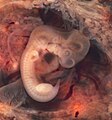 Human Embryo (7th week of pregnancy) (304334264).jpg 1,874 × 2,000; 1.36 MB
-
 Human Yolk Sac from Tubal Pregnancy (32847892157).jpg 1,946 × 1,887; 891 KB
-
 Human Yolk Sac from Tubal Pregnancy (33914344418).jpg 1,670 × 2,559; 676 KB
-
 Human Yolk Sac from Tubal Pregnancy (33914344548).jpg 1,771 × 2,516; 1.06 MB
-
 Human Yolk Sac from Tubal Pregnancy (33914345538).jpg 1,643 × 2,167; 669 KB
-
 Human Yolk Sac from Tubal Pregnancy (47002175724).jpg 2,096 × 2,274; 1.02 MB
-
 Human Yolk Sac from Tubal Pregnancy (47002176474).jpg 2,026 × 1,962; 1.16 MB
-
 Human Yolk Sac from Tubal Pregnancy (47739058042).jpg 1,744 × 2,647; 1.35 MB
-
 Human Yolk Sac from Tubal Pregnancy (47739058172).jpg 2,330 × 2,384; 1.55 MB
-
 Human Yolk Sac from Tubal Pregnancy (47791378311).jpg 1,514 × 2,075; 527 KB
-
 Hypersegmented Neutrophils (36831145373).jpg 960 × 690; 64 KB
-
 Infectious Mononucleosis. Peripheral Blood, Wright Stain (25116997017).jpg 1,619 × 1,760; 816 KB
-
 Inhibin (focal +) (2164277304).jpg 871 × 751; 564 KB
-
 Intraductal Papilloma of the Breast (24808837466).jpg 2,048 × 1,536; 668 KB
-
 Intramuscular Lipoma (38578598922).jpg 1,814 × 1,673; 994 KB
-
 Intramuscular Lipoma (51050168853).jpg 2,197 × 1,812; 979 KB
-
 Intramuscular Lipoma (51050168908).jpg 2,247 × 2,006; 1.86 MB
-
 Intramuscular Lipoma (51050979822).jpg 2,243 × 1,927; 1.53 MB
-
 Invasive Ductal Carcinoma of the Breast (39783804110).jpg 2,942 × 1,902; 3.1 MB
-
 Invasive Lobular Carcinoma (43256345075).jpg 2,132 × 2,164; 1.17 MB
-
 Invasive Lobular Carcinoma (44139478902).jpg 2,204 × 2,034; 1.4 MB
-
 Invasive Lobular Carcinoma (44187539911).jpg 2,663 × 1,426; 1.53 MB
-
 Invasive Lobular Carcinoma of the Breast (33768522968).jpg 1,405 × 705; 156 KB
-
 Invasive Lobular Carcinoma, CAM 5.2 Low Molecular Weight Keratin (43256344145).jpg 2,107 × 1,949; 1.04 MB
-
 Invasive Lobular Carcinoma, Estrogen Receptor (42352322550).jpg 2,197 × 2,027; 748 KB
-
 Involucrum (Extracortical New Bone Formation) in Chronic Osteomyelitis (48327308712).jpg 2,452 × 1,762; 1.54 MB
-
 Juvenile Granulosa Cell Tumor, Ovary (25394519147).jpg 2,333 × 2,030; 1.89 MB
-
 Juvenile Granulosa Cell Tumor, Ovary (25394681457).jpg 2,455 × 2,191; 2.75 MB
-
 Juvenile Granulosa Cell Tumor, Ovary (26392980378).jpg 1,976 × 2,269; 1.95 MB
-
 Juvenile Granulosa Cell Tumor, Ovary (26393564228).jpg 2,367 × 1,835; 2.32 MB
-
 Juvenile Granulosa Cell Tumor, Ovary (38455669850).jpg 2,067 × 1,682; 1.83 MB
.jpg)
.jpg)
.jpg)
.jpg)
.jpg)
.jpg)
.jpg)
.jpg)
.jpg)
.jpg)
.jpg)
.jpg)
.jpg)
.jpg)
.jpg)
_Pigment_(17509353414).jpg)
.jpg)
.jpg)
.jpg)
.jpg)
.jpg)
.jpg)
.jpg)
.jpg)
.jpg)
.jpg)
.jpg)
.jpg)
.jpg)
_(2164275352).jpg)
_(2164276824).jpg)
.jpg)
_(2163477587).jpg)
_(2164277470).jpg)
.jpg)
.jpg)
.jpg)
.jpg)
.jpg)
.jpg)
.jpg)
.jpg)
.jpg)
.jpg)
.jpg)
.jpg)
.jpg)
.jpg)
_NZ7_1386_(51440373932).jpg)
_NZ7_1388_(51441869729).jpg)
_NZ7_1389_(51441868574).jpg)
.jpg)
.jpg)
.jpg)
.jpg)
.jpg)
.jpg)
.jpg)
.jpg)
.jpg)
.jpg)
.jpg)
.jpg)
.jpg)
.jpg)
.jpg)
.jpg)
.jpg)
.jpg)
.jpg)
.jpg)
.jpg)
.jpg)
.jpg)
.jpg)
.jpg)
.jpg)
.jpg)
.jpg)
.jpg)
_(2163477507).jpg)
_(2163477293).jpg)
.jpg)
.jpg)
.jpg)
.jpg)
.jpg)
.jpg)
.jpg)
_(21739375573).jpg)
.jpg)
.jpg)
.jpg)
.jpg)
.jpg)
.jpg)
.jpg)
.jpg)
.jpg)
_(46262991251).jpg)
.jpg)
_(2164277240).jpg)
.jpg)
.jpg)
_(4837907128).jpg)
_(4837290693).jpg)
_(4837292483).jpg)
_(4837293823).jpg)
_(4837906214).jpg)
.jpg)
.jpg)
.jpg)
.jpg)
.jpg)
.jpg)
.jpg)
.jpg)
.jpg)
_(48879295668).jpg)
_(48879851431).jpg)
_(48880043332).jpg)
.jpg)
.jpg)
.jpg)
_(5241534941).jpg)
_(5241533387).jpg)
_(5241535707).jpg)
.jpg)
.jpg)
.jpg)
.jpg)
.jpg)
.jpg)
.jpg)
.jpg)
.jpg)
.jpg)
.jpg)
.jpg)
.jpg)
.jpg)
.jpg)
.jpg)
.jpg)
.jpg)
.jpg)
.jpg)
.jpg)
_(2164277392).jpg)
.jpg)
_of_the_Cervix_(44001403165).jpg)
_(304334264).jpg)
.jpg)
.jpg)
.jpg)
.jpg)
.jpg)
.jpg)
.jpg)
.jpg)
.jpg)
.jpg)
.jpg)
_(2164277304).jpg)
.jpg)
.jpg)
.jpg)
.jpg)
.jpg)
.jpg)
.jpg)
.jpg)
.jpg)
.jpg)
.jpg)
.jpg)
_in_Chronic_Osteomyelitis_(48327308712).jpg)
.jpg)
.jpg)
.jpg)
.jpg)
.jpg)
.jpg)
.jpg)
.jpg)
.jpg)
.jpg)
.jpg)
.jpg)
.jpg)
.jpg)
.jpg)
.jpg)
.jpg)
.jpg)
.jpg)
.jpg)
.jpg)
.jpg)
_(26393361318).jpg)