Category:Fasciola hepatica
Jump to navigation
Jump to search
parasitic flatworm in mammal livers | |||||||||||||||||||||||||||||
| Upload media | |||||||||||||||||||||||||||||
| Instance of | |||||||||||||||||||||||||||||
|---|---|---|---|---|---|---|---|---|---|---|---|---|---|---|---|---|---|---|---|---|---|---|---|---|---|---|---|---|---|
| Different from | |||||||||||||||||||||||||||||
| |||||||||||||||||||||||||||||
| |||||||||||||||||||||||||||||
| Taxon author | Carl Linnaeus, 1758 | ||||||||||||||||||||||||||||
| |||||||||||||||||||||||||||||
Fasciola hepatica (the common liver fluke) is one of the most important parasite of sheep and cattle.
Subcategories
This category has the following 3 subcategories, out of 3 total.
F
- Fascioliasis (9 F)
I
Media in category "Fasciola hepatica"
The following 64 files are in this category, out of 64 total.
-
Dewe vatche botaye-fritche.jpeg 640 × 480; 80 KB
-
EB1911 Trematodes - Fasciola hepatica.jpg 773 × 1,047; 344 KB
-
EB1911 Trematodes - Five stages in the life-history of Fasciola hepatica.jpg 720 × 1,029; 303 KB
-
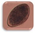
Egg of Fasciola hepatica 08G0041 lores.jpg 531 × 392; 48 KB
-
Egg of Liver Fluke.jpg 3,264 × 2,448; 1.22 MB
-
F. hepatica adults in bile duct.jpg 342 × 256; 27 KB
-
F. hepatica cercariae.webm 5.9 s, 1,920 × 1,080; 2.25 MB
-
F. hepatica hypertrophia of bile duct.jpg 288 × 216; 25 KB
-
F.magna 1 a F.hepatica 2.jpg 336 × 336; 16 KB
-
Falsciola hepática en Conducto Biliar..jpg 1,936 × 2,592; 2.12 MB
-
Fasciola hepatica (01).png 1,232 × 1,259; 553 KB
-
Fasciola hepatica (Eier).jpg 1,000 × 640; 163 KB
-
Fasciola hepatica (Körpermitte, quer, Teilausschnitt).jpg 2,592 × 1,790; 1.37 MB
-
Fasciola hepatica (Linnaeus, 1758) 2013 000-2.jpg 6,073 × 2,500; 5.54 MB
-
Fasciola hepatica (Linnaeus, 1758) 2013 000.jpg 14,980 × 6,166; 7.3 MB
-
Fasciola hepatica (Linnaeus, 1758) 2013 001-2.jpg 7,311 × 3,000; 6.53 MB
-
Fasciola hepatica (Linnaeus, 1758) 2013 001.jpg 14,816 × 6,080; 11.71 MB
-
Fasciola hepatica 01 Pengo.jpg 1,600 × 1,200; 456 KB
-
Fasciola hepatica adult (01).png 1,499 × 1,684; 663 KB
-
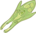
Fasciola hepatica cercaria (01).png 1,093 × 1,154; 259 KB
-
Fasciola hepatica egg (01).png 851 × 931; 190 KB
-
Fasciola Hepatica egg (liverfluke) (15767790812).jpg 129 × 123; 4 KB
-
Fasciola hepatica egg embryonated (01).png 812 × 977; 193 KB
-
Fasciola Hepatica Ei.png 169 × 261; 117 KB
-
Fasciola hepatica metacercaria (01).png 1,062 × 1,207; 272 KB
-
Fasciola hepatica prevalence.jpg 1,425 × 625; 504 KB
-
Fasciola hepatica rediae (01).png 1,364 × 1,474; 400 KB
-
Fasciola hepatica sporocyte (01).png 1,385 × 1,378; 339 KB
-
Fasciola hepatica wm (3) anterior.jpg 1,280 × 720; 50 KB
-
Fasciola hepatica wm (5) anterior with eggs.jpg 1,280 × 720; 140 KB
-
Fasciola hepatica wm anterior.jpg 1,280 × 720; 74 KB
-
Fasciola hepatica wm posteior.jpg 1,280 × 720; 101 KB
-
Fasciola hepatica wm uterus, ceca and yolk glands.jpg 1,280 × 720; 120 KB
-
Fasciola hepatica wm with eggs.jpg 1,280 × 720; 141 KB
-
Fasciola hepatica wm.jpg 1,280 × 720; 221 KB
-
Fasciola hepatica x 100mag (1) (7686932610).jpg 1,024 × 768; 172 KB
-
Fasciola hepatica x 100mag (2) (7686932964).jpg 1,024 × 768; 170 KB
-
Fasciola hepatica x 400mag (7686933314).jpg 1,024 × 768; 174 KB
-
Fasciola hepatica.JPG 448 × 336; 39 KB
-
Fasciola hepatica.jpg 631 × 270; 19 KB
-
Fasciola hepatica2.jpg 631 × 270; 112 KB
-
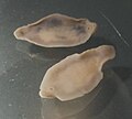
Fasciola-hepatica-adults.jpg 1,398 × 681; 166 KB
-
Fasciola-hepatica-bileduct.jpg 1,827 × 2,371; 484 KB
-
FasciolaHepatica1.jpg 1,201 × 1,608; 767 KB
-
Fhepatica cercariae movement.gif 640 × 592; 2.24 MB
-
Foete dewe.jpg 640 × 360; 50 KB
-
Foetite grangreneuse foete atnaedjes.JPG 640 × 360; 123 KB
-
Hatching of F. hepatica miracidia.ogv 1 min 18 s, 1,280 × 720; 5.41 MB
-
Huevos de duelas hepáticas comparación.jpg 3,000 × 4,000; 510 KB
-
Infectiology - Fasciola hepatica - Metacercaria -- Smart-Servier.png 617 × 636; 95 KB
-
Infectiology - Fasciola hepatica - Redia -- Smart-Servier.png 691 × 635; 106 KB
-
Meyers b16 s0768a.jpg 800 × 630; 131 KB
-
Nema Fig5.gif 650 × 393; 79 KB
-
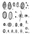
Parasitic worm eggs.tif 1,438 × 1,517; 1 MB
-
PSM V23 D761 The liver fluke fasciola hepatica.jpg 558 × 1,320; 149 KB
-
PSM V23 D762 Egg of the liver fluke.jpg 1,465 × 770; 146 KB
-
PSM V23 D762 Fully developed fluke egg and the cilia for water propulsion.jpg 1,528 × 1,357; 403 KB
-
PSM V23 D763 The trematode becomes a sporocyst in a snail.jpg 1,609 × 999; 285 KB
-
PSM V23 D764 Young redia forming into liver fluke.jpg 795 × 1,786; 298 KB
-
PSM V23 D765 Free cercaria in water and cysts attached to the grass.jpg 1,559 × 1,176; 142 KB
-
Saguaype.jpg 1,061 × 957; 125 KB
-
Syncytial epithelia.jpg 525 × 346; 37 KB
-
TrematodesFig8 EncBrit1911.png 1,100 × 1,824; 189 KB


