Category:Diagnostic radiology
Jump to navigation
Jump to search
Wikimedia category | |||||
| Upload media | |||||
| Instance of | |||||
|---|---|---|---|---|---|
| |||||
Media in category "Diagnostic radiology"
The following 200 files are in this category, out of 259 total.
(previous page) (next page)-
-
-
-
-
-
-
-
-
-
A-Novel-Immunofluorescent-Computed-Tomography-(ICT)-Method-to-Localise-and-Quantify-Multiple-pone.0053245.s001.ogv 1 min 0 s, 1,104 × 480; 17.79 MB
-
A-Novel-Immunofluorescent-Computed-Tomography-(ICT)-Method-to-Localise-and-Quantify-Multiple-pone.0053245.s002.ogv 1 min 0 s, 1,104 × 480; 7.34 MB
-
A-Novel-Immunofluorescent-Computed-Tomography-(ICT)-Method-to-Localise-and-Quantify-Multiple-pone.0053245.s003.ogv 1 min 0 s, 1,104 × 480; 1.96 MB
-
-
-
-
-
A-Physiologically-Motivated-Compartment-Based-Model-of-the-Effect-of-Inhaled-Hypertonic-Saline-on-pone.0111972.s006.ogv 1.0 s, 1,392 × 1,040; 1.65 MB
-
-
-
-
-
-
-
-
-
-
Augmented-Endoscopic-Images-Overlaying-Shape-Changes-in-Bone-Cutting-Procedures-pone.0161815.s002.ogv 2 min 27 s, 480 × 424; 1.93 MB
-
-
-
-
-
-
Chronic-High-Fat-Diet-Impairs-Collecting-Lymphatic-Vessel-Function-in-Mice-pone.0094713.s003.ogv 18 s, 512 × 512; 230 KB
-
Chronic-High-Fat-Diet-Impairs-Collecting-Lymphatic-Vessel-Function-in-Mice-pone.0094713.s004.ogv 18 s, 512 × 512; 291 KB
-
Chronic-High-Fat-Diet-Impairs-Collecting-Lymphatic-Vessel-Function-in-Mice-pone.0094713.s005.ogv 14 s, 512 × 512; 110 KB
-
Chronic-High-Fat-Diet-Impairs-Collecting-Lymphatic-Vessel-Function-in-Mice-pone.0094713.s006.ogv 14 s, 512 × 512; 117 KB
-
-
-
-
-
-
-
Comparison-of-Sum-Absolute-QRST-Integral-and-Temporal-Variability-in-Depolarization-and-pone.0057175.s001.ogv 1 min 31 s, 616 × 616; 18.57 MB
-
Comparison-of-Sum-Absolute-QRST-Integral-and-Temporal-Variability-in-Depolarization-and-pone.0057175.s002.ogv 1 min 14 s, 616 × 616; 17.56 MB
-
Correlative-Nanoscale-3D-Imaging-of-Structure-and-Composition-in-Extended-Objects-pone.0050124.s002.ogv 1 min 2 s, 464 × 384; 6.83 MB
-
Creating-High-Resolution-Multiscale-Maps-of-Human-Tissue-Using-Multi-beam-SEM-pcbi.1005217.s004.ogv 5.0 s, 1,000 × 1,149; 5.71 MB
-
Creating-High-Resolution-Multiscale-Maps-of-Human-Tissue-Using-Multi-beam-SEM-pcbi.1005217.s005.ogv 4.2 s, 742 × 980; 12.66 MB
-
Creating-High-Resolution-Multiscale-Maps-of-Human-Tissue-Using-Multi-beam-SEM-pcbi.1005217.s006.ogv 5.0 s, 750 × 652; 24.87 MB
-
-
-
-
-
-
-
Deep-Learning-Automates-the-Quantitative-Analysis-of-Individual-Cells-in-Live-Cell-Imaging-pcbi.1005177.s021.ogv 6.4 s, 1,220 × 1,020; 2.13 MB
-
Deep-Learning-Automates-the-Quantitative-Analysis-of-Individual-Cells-in-Live-Cell-Imaging-pcbi.1005177.s022.ogv 6.4 s, 1,280 × 1,080; 5.76 MB
-
Deep-Learning-Automates-the-Quantitative-Analysis-of-Individual-Cells-in-Live-Cell-Imaging-pcbi.1005177.s023.ogv 6.4 s, 1,220 × 1,020; 2.16 MB
-
Deep-Learning-Automates-the-Quantitative-Analysis-of-Individual-Cells-in-Live-Cell-Imaging-pcbi.1005177.s024.ogv 6.4 s, 1,220 × 1,020; 1.16 MB
-
Deep-Learning-Automates-the-Quantitative-Analysis-of-Individual-Cells-in-Live-Cell-Imaging-pcbi.1005177.s025.ogv 6.4 s, 1,280 × 1,080; 11.76 MB
-
-
-
-
-
Dynamic-Characterization-of-the-CT-Angiographic-Spot-Sign-pone.0090431.s001.ogv 4.6 s, 512 × 512; 307 KB
-
Dynamic-Characterization-of-the-CT-Angiographic-Spot-Sign-pone.0090431.s002.ogv 4.6 s, 512 × 512; 473 KB
-
-
-
-
-
-
Endoscopic-Gold-Fiducial-Marker-Placement-into-the-Bladder-Wall-to-Optimize-Radiotherapy-Targeting-pone.0089754.s001.ogv 2 min 50 s, 960 × 540; 2.58 MB
-
-
-
-
-
FISICO-Fast-Image-SegmentatIon-COrrection-pone.0156035.s001.ogv 1 min 47 s, 712 × 480; 12.91 MB
-
Gas-Filled-Phospholipid-Nanoparticles-Conjugated-with-Gadolinium-Play-a-Role-as-a-Potential-pone.0034333.s001.ogv 1 min 5 s, 480 × 272; 2.29 MB
-
Gas-Filled-Phospholipid-Nanoparticles-Conjugated-with-Gadolinium-Play-a-Role-as-a-Potential-pone.0034333.s002.ogv 1 min 17 s, 480 × 272; 2.18 MB
-
Gas-Filled-Phospholipid-Nanoparticles-Conjugated-with-Gadolinium-Play-a-Role-as-a-Potential-pone.0034333.s003.ogv 1 min 4 s, 480 × 272; 3.44 MB
-
Gas-Filled-Phospholipid-Nanoparticles-Conjugated-with-Gadolinium-Play-a-Role-as-a-Potential-pone.0034333.s004.ogv 1 min 4 s, 480 × 272; 5.71 MB
-
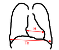
Herz-thorax-quotient a.png 1,468 × 1,251; 19 KB
-
Herz-thorax-quotient b.png 1,468 × 1,251; 21 KB
-
-
-
-
-
How-Do-You-Feel-when-You-Cant-Feel-Your-Body-Interoception-Functional-Connectivity-and-Emotional-pone.0098769.s007.ogv 1 min 40 s, 640 × 480; 2.34 MB
-
-
In-Amnio-MRI-of-Mouse-Embryos-pone.0109143.s002.ogv 24 s, 720 × 576; 3.73 MB
-
In-Amnio-MRI-of-Mouse-Embryos-pone.0109143.s003.ogv 24 s, 720 × 576; 3.08 MB
-
In-Amnio-MRI-of-Mouse-Embryos-pone.0109143.s004.ogv 24 s, 720 × 576; 3.95 MB
-
-
-
-
-
-
-
-
-
-
-
-
-
-
-
-
-
-
-
-
-
-
Increased-Echogenicity-and-Radiodense-Foci-on-Echocardiogram-and-MicroCT-in-Murine-Myocarditis-pone.0159971.s012.ogv 1 min 7 s, 340 × 480; 7.1 MB
-
-
-
-
-
Inside-Out-Modern-Imaging-Techniques-to-Reveal-Animal-Anatomy-pone.0017879.s001.ogv 1 min 0 s, 736 × 480; 3.1 MB
-
Inside-Out-Modern-Imaging-Techniques-to-Reveal-Animal-Anatomy-pone.0017879.s002.ogv 53 s, 1,920 × 1,080; 44.99 MB
-
Inside-Out-Modern-Imaging-Techniques-to-Reveal-Animal-Anatomy-pone.0017879.s003.ogv 1 min 0 s, 736 × 480; 7.39 MB
-
Inside-Out-Modern-Imaging-Techniques-to-Reveal-Animal-Anatomy-pone.0017879.s004.ogv 1 min 0 s, 736 × 480; 12.83 MB
-
Inside-Out-Modern-Imaging-Techniques-to-Reveal-Animal-Anatomy-pone.0017879.s005.ogv 2 min 0 s, 736 × 480; 41.05 MB
-
Investigation-of-6-18F-Fluoromaltose-as-a-Novel-PET-Tracer-for-Imaging-Bacterial-Infection-pone.0107951.s002.ogv 15 s, 1,600 × 1,088; 4.46 MB
-
LaminiticRadiographicMeasurements.jpg 800 × 578; 50 KB
-
-
-
-
-
-
-
-
-
-
-
-
-
-
Mechanisms-of-Hearing-Loss-after-Blast-Injury-to-the-Ear-pone.0067618.s002.ogv 2 min 0 s, 640 × 480; 10.52 MB
-
Minimum-Field-Strength-Simulator-for-Proton-Density-Weighted-MRI-pone.0154711.s001.ogv 10 s, 288 × 120; 597 KB
-
-
-
-
-
-
-
-
-
-
-
Mouse-Model-of-Lymph-Node-Metastasis-via-Afferent-Lymphatic-Vessels-for-Development-of-Imaging-pone.0055797.s002.ogv 1 min 16 s, 768 × 576; 1.83 MB
-
-
-
-
MRI-Based-Localisation-and-Quantification-of-Abscesses-following-Experimental-S.-aureus-Intravenous-pone.0154705.s008.ogv 3 min 26 s, 504 × 504; 22.19 MB
-
-
-
-
-
Neural-Substrates-for-the-Motivational-Regulation-of-Motor-Recovery-after-Spinal-Cord-Injury-pone.0024854.s001.ogv 1 min 18 s, 320 × 240; 3.02 MB
-
-
-
-
-
-
-
-
-
-
-
Open-Source-Selective-Laser-Sintering-(OpenSLS)-of-Nylon-and-Biocompatible-Polycaprolactone-pone.0147399.s006.ogv 34 s, 1,920 × 1,080; 23.17 MB
-
-
-
-
-
-
-
-
-
-
-
RM de abdomen con gadolinio.jpg 284 × 363; 43 KB
-
-
-
-
-
-
Röntgenologisch onderzoek van de bevolking van Boekel-517526.ogv 1 min 56 s, 384 × 288; 10.04 MB
-
-
-
-
-
-
-
-
-
Seeing-through-Musculoskeletal-Tissues-Improving-In-Situ-Imaging-of-Bone-and-the-Lacunar-pone.0150268.s010.ogv 19 s, 1,024 × 1,024; 1.92 MB
-
Seeing-through-Musculoskeletal-Tissues-Improving-In-Situ-Imaging-of-Bone-and-the-Lacunar-pone.0150268.s011.ogv 13 s, 1,024 × 1,024; 1.67 MB
-
Shared-Brain-Lateralization-Patterns-in-Language-and-Acheulean-Stone-Tool-Production-A-Functional-pone.0072693.s001.ogv 46 s, 1,920 × 1,080; 15.08 MB
-
-


