Category:CT images of cancers of bronchus and lung
Jump to navigation
Jump to search
Subcategories
This category has the following 6 subcategories, out of 6 total.
Media in category "CT images of cancers of bronchus and lung"
The following 23 files are in this category, out of 23 total.
-
 Adenocarcinoma - CT scan (5499628365).jpg 512 × 408; 43 KB
-
 Biopsie Lunge Computertomographie BC.png 1,190 × 948; 572 KB
-
 CT of a lung nodule abutting a cystic airspace.png 126 × 124; 17 KB
-
 CT of a lung nodule with pleural retraction.png 161 × 156; 30 KB
-
 CT of a subpleural nodule with pleural retraction.png 152 × 166; 23 KB
-
 CT of ground glass lung nodule.png 342 × 256; 98 KB
-
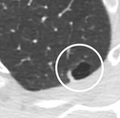
 CT of lung nodule with bubble-like lucencies.png 160 × 139; 29 KB
-
 CT of lung nodule with smooth border.png 272 × 232; 58 KB
-
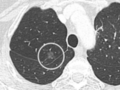
 CT of lung nodule with vascular convergence (crop).png 306 × 268; 94 KB
-
 CT of lung nodule with vascular convergence.png 307 × 514; 186 KB
-
 CT of part solid lung nodule.png 396 × 354; 154 KB
-
 CT of spiculated lung nodule with bubble-like lucencies.png 145 × 184; 31 KB
-
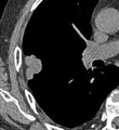
 CT scan of lung cancer with cavitation.png 495 × 392; 173 KB
-
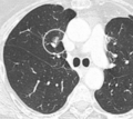
 EmphysemaPlusLungCA.png 810 × 810; 327 KB
-
 EmphysemaPlusLungCAMark.png 810 × 810; 324 KB
-
 LungcaCT.PNG 1,178 × 869; 765 KB
-
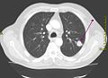
 Pancoast Tumor 2.jpg 640 × 345; 46 KB
-

-
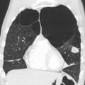
 Thorax CT cor MPR peripheres Bronchialcarcinom li OF.jpg 301 × 277; 9 KB
-
 Thorax CT pcor Cut 3D Volume Rendering Haut.jpg 326 × 276; 13 KB
-
 Thorax CT peripheres Brronchialcarcinom li OF.jpg 506 × 366; 14 KB
-
 Tumor in L. lung-3D CT volume rendering.png 818 × 716; 1.27 MB
-
 Гамартома КТ.jpg 724 × 777; 156 KB
.jpg)


.png)


.jpg)


