Category:Base pairing
Jump to navigation
Jump to search
unit consisting of two nucleobases bound to each other by hydrogen bonds: either adenine–thymine or guanine–cytosine in natural DNA (additional types occur in RNA) | |||||
| Upload media | |||||
| Instance of | |||||
|---|---|---|---|---|---|
| Measured physical quantity |
| ||||
| |||||
Subcategories
This category has the following 2 subcategories, out of 2 total.
- SVG base pairing (59 F)
B
- Base pair mismatch (13 F)
Media in category "Base pairing"
The following 103 files are in this category, out of 103 total.
-
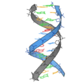
1bna naba ribbon v.png 3,228 × 3,180; 513 KB
-
6оо4о54п5.tif 759 × 1,044; 143 KB
-
A and yT.png 517 × 292; 13 KB
-
A bonded to xT.png 469 × 230; 10 KB
-
Abasic-pivot-substitution-harnesses-target-specificity-of-RNA-interference-ncomms10154-s2.ogv 41 s, 320 × 240; 2.46 MB
-
Abasic-pivot-substitution-harnesses-target-specificity-of-RNA-interference-ncomms10154-s3.ogv 41 s, 320 × 240; 3.16 MB
-
Abasic-pivot-substitution-harnesses-target-specificity-of-RNA-interference-ncomms10154-s4.ogv 41 s, 320 × 240; 2.27 MB
-
Adenine-Cytosine base pairing.svg 726 × 450; 25 KB
-
AGCT DNA mini.png 257 × 259; 9 KB
-
AT base pair jypx3.png 588 × 404; 11 KB
-
AT DNA base pair tr.png 559 × 323; 19 KB
-
AT-DNA-base-pair-3D-vdW.png 1,292 × 780; 131 KB
-
AT-GC.jpg 835 × 458; 31 KB
-
AUreverse.png 372 × 223; 37 KB
-
B&Z&A DNA formula.sr.png 849 × 573; 156 KB
-
Base Scheme.jpg 623 × 636; 157 KB
-
Blausen 0321 DNA 1.png 2,600 × 4,600; 3.25 MB
-
Blocosadnbyaal.JPG 956 × 621; 93 KB
-
C-C pairing.pdf 352 × 387; 8 KB
-
C-C pairing.svg 225 × 248; 19 KB
-
Chargaffs regel 2.jpg 541 × 185; 68 KB
-
Chargaffs regel.jpg 541 × 185; 67 KB
-
Cis and Trans orientation of glycosidic bonds.png 1,014 × 1,002; 125 KB
-
Cis Trans orientations of glycosidic Bond.png 972 × 996; 120 KB
-
Cytosin-Guanine base pairing.svg 765 × 522; 32 KB
-
Cytosine tautomerism.svg 963 × 494; 30 KB
-
Cytosine-Hypoxanthine Base Pairing.png 1,024 × 768; 37 KB
-
Diagram showing a double helix of a chromosome CRUK 065-hi.png 301 × 388; 46 KB
-
DNA Diagram.png 336 × 192; 29 KB
-
Dna strand.png 432 × 507; 50 KB
-
DNA Structure+Key+Labelled pn NoBB.sr.png 1,037 × 977; 553 KB
-
DNA Structure+Key+Labelled Swetransl.png 3,075 × 3,000; 3.43 MB
-
DNA Structure+Key+Labelled-es.png 3,075 × 3,000; 3.28 MB
-
DNA Structure+Key+Labelled.pn NoBB ar.png 3,075 × 3,000; 3.1 MB
-
DNA Structure+Key+Labelled.pn NoBB cs.png 3,075 × 3,000; 3.22 MB
-
DNA Structure+Key+Labelled.pn NoBB gl.png 3,075 × 3,000; 4.28 MB
-
DNA Structure+Key+Labelled.pn NoBB nl.svg 640 × 600; 5.32 MB
-
DNA Structure+Key+Labelled.pn NoBB.png 3,024 × 3,000; 2.64 MB
-
DNA Structure+Key+Labelled.png 3,075 × 3,000; 4.69 MB
-
DNA structuur1 200.png 200 × 368; 8 KB
-
DNA yapısı.png 3,075 × 3,000; 3.81 MB
-
DNA-internal-CpG-dinucleotide.png 1,134 × 1,134; 29 KB
-
DNA-labels.png 2,048 × 1,969; 97 KB
-
Dna-split.png 202 × 397; 12 KB
-
DNA-structure-composition-pt-br.png 817 × 408; 123 KB
-
Figure1New.png 960 × 720; 123 KB
-
GC base pair jypx3.png 593 × 436; 12 KB
-
GC DNA base pair tr.png 558 × 345; 21 KB
-
GC DNA base pair.png 554 × 459; 18 KB
-
GC DNA base pair.sr.svg 638 × 401; 11 KB
-
GC Watson Crick basepair.png 354 × 241; 3 KB
-
GC-DNA-base-pair-3D-vdW.png 2,016 × 832; 228 KB
-
GCreverse.png 389 × 325; 47 KB
-
-
-
-
-
Guanine tautomerism.svg 1,235 × 492; 39 KB
-
Guanine-Adenine base pairing.svg 798 × 465; 30 KB
-
Guanine-Guanine base pairing.svg 903 × 405; 28 KB
-
H bond in RNA translation.jpg 378 × 394; 36 KB
-
H-DNA Triads.jpg 1,178 × 740; 109 KB
-
Hoogsteen Watson Crick pairing-en.svg 512 × 288; 36 KB
-
Identification of inosine.png 503 × 565; 71 KB
-
Interacao das bases nitrogenadas.png 1,387 × 734; 77 KB
-
Isoguanin-Isocytosin-Basenpaarung V1.svg 403 × 378; 49 KB
-
Isoguanine-Isocytosine-Base pair V2.svg 403 × 336; 44 KB
-
Miri10.png 620 × 369; 18 KB
-
Molnupiravir AU bonding.svg 512 × 283; 7 KB
-
Molnupiravir GC bonding.svg 512 × 298; 8 KB
-
Non-canonical base pairing Fig1a.png 1,209 × 584; 25 KB
-
Non-canonical base pairing Fig1b.png 1,189 × 777; 31 KB
-
Non-canonical base pairing Fig3.png 431 × 378; 122 KB
-
Non-canonical base pairing Fig4a.png 420 × 450; 28 KB
-
Non-canonical base pairing Fig4b.png 420 × 450; 36 KB
-
Non-canonical base pairing Fig5.png 771 × 144; 28 KB
-
Nucleótido.png 268 × 388; 12 KB
-
Paire de base.gif 330 × 476; 15 KB
-
Palindrom.PNG 169 × 104; 10 KB
-
Par bases watson crick reverse gl.png 1,123 × 794; 49 KB
-
Par bases watson crick reverse.png 1,123 × 794; 45 KB
-
Par bases watson-crick.png 1,123 × 794; 44 KB
-
Pencil sketch of molecular diagram Wellcome L0051226.jpg 4,924 × 6,261; 3.7 MB
-
Pencil sketch of the DNA double helix by Francis Crick Wellcome L0033046.jpg 3,698 × 4,736; 1,002 KB
-
Pencil sketch of the DNA double helix by Francis Crick Wellcome L0051225.jpg 4,900 × 6,254; 3.88 MB
-
Poly I-C structure de.svg 415 × 285; 53 KB
-
Poly I-C structure.svg 421 × 304; 143 KB
-
RNA Base Pairings.png 744 × 183; 34 KB
-
RNA Wobble Base Pair.png 355 × 299; 20 KB
-
RNA-comparedto-DNA thymineAndUracilCorrected.png 530 × 538; 78 KB
-
TA*T and CG*C+ Hoogsteen Binding in Triplex DNA.tif 2,048 × 700; 187 KB
-
TABASCO-A-single-molecule-base-pair-resolved-gene-expression-simulator-1471-2105-8-480-S1.ogv 30 s, 1,024 × 768; 6.24 MB
-
Thymin Keto-Enol-Tautomerie.svg 1,149 × 485; 31 KB
-
Thymine-Adenine base pairing.svg 720 × 444; 24 KB
-
Thymine-enol-Guanine base pairing.svg 741 × 510; 31 KB
-
TYP DNA.JPG 772 × 620; 28 KB
-
TYP RNA.JPG 772 × 620; 29 KB
-
Vrevwr33.tif 476 × 237; 9 KB
-
WobbleBasePairs.jpg 259 × 368; 45 KB
-
WobbleBasePairsUracil.jpg 387 × 473; 39 KB
-
Рис.GC.jpg 2,128 × 3,068; 398 KB
-
Универсальные основания.png 493 × 387; 36 KB

