Category:Anatomy of the human muscular system
Jump to navigation
Jump to search
Subcategories
This category has the following 14 subcategories, out of 14 total.
Media in category "Anatomy of the human muscular system"
The following 161 files are in this category, out of 161 total.
-
(Muscles of the human body) (4647096651).jpg 1,732 × 2,730; 600 KB
-
A human hand anatomised and preserved Wellcome L0036404.jpg 3,008 × 1,960; 727 KB
-
A male écorché figure by Giulio Bonasone.jpg 295 × 384; 36 KB
-
Abdomen muscle.png 1,149 × 1,351; 846 KB
-
Albinus skeleton w muscles.jpg 1,134 × 1,560; 685 KB
-
An écorché; with left arm extended to the side, seen from th Wellcome V0007962EL.jpg 1,080 × 1,656; 1.24 MB
-
Anatomia esterna de corporumano . . . Wellcome L0009624.jpg 1,124 × 1,658; 905 KB
-
Anatomia esterna de corporumano . . . Wellcome L0009625.jpg 1,108 × 1,640; 809 KB
-
Anatomia esterna de corporumano . . . Wellcome L0009626.jpg 1,120 × 1,684; 847 KB
-
Anatomia esterna de corporumano . . . Wellcome L0009627.jpg 1,128 × 1,674; 826 KB
-
Anatomia esterna de corporumano . . . Wellcome L0009628.jpg 1,137 × 1,669; 806 KB
-
Anatomia esterna del corpo umano, title page. Wellcome L0023846.jpg 1,204 × 1,666; 741 KB
-
Anatomia esterna del corpo umano, title page. Wellcome L0023847.jpg 1,174 × 1,636; 746 KB
-
Anatomia esterna del corpo umano, title page. Wellcome L0023848.jpg 1,180 × 1,616; 754 KB
-
Anatomia esterna del corpo umano, title page. Wellcome L0040365.jpg 2,792 × 4,000; 2.01 MB
-
Anatomia esterna del corpo umano, title page. Wellcome L0040366.jpg 2,776 × 4,072; 2.19 MB
-
Anatomical figure displaying the muscles of the torso Wellcome L0040034.jpg 2,604 × 3,948; 2.84 MB
-
Anatomical figure. Etching by A. Cattani, 1780-1781, after Wellcome L0027191.jpg 750 × 2,477; 884 KB
-
Anatomical figure. Etching by A. Cattani, 1781, after E. Wellcome L0027190.jpg 750 × 2,470; 862 KB
-
Anatomical sketches after Valverde; muscles. Wellcome L0011864.jpg 1,556 × 1,274; 1.02 MB
-
Anatomy posture and body mechanics 08.jpg 653 × 1,024; 244 KB
-
Anatomy, physiology and laws of health; (1885) (14594982848).jpg 1,320 × 2,860; 963 KB
-
Anatomy, skeletons of two foetuses, two and Wellcome V0007790.jpg 648 × 486; 83 KB
-
Archiv für Anatomie, Physiologie und wissenschaftliche Medicin (1865) (20319181912).jpg 2,976 × 2,226; 1.09 MB
-
Avant bras.png 2,381 × 2,938; 2.64 MB
-
Bell heads with neck muscles.jpg 791 × 581; 231 KB
-
Bougle top02.jpg 1,200 × 1,980; 320 KB
-
Bougle top04.jpg 1,200 × 2,033; 396 KB
-
Bougle whole2 retouched.png 1,014 × 3,006; 3.16 MB
-
Bougle whole2.jpg 1,000 × 3,012; 309 KB
-
Bougle whole5.jpg 1,000 × 3,004; 620 KB
-
Braus 1921 115.png 1,620 × 1,584; 7.36 MB
-
Braus 1921 120.png 1,640 × 2,632; 12.37 MB
-
Braus 1921 131.png 1,805 × 2,655; 13.74 MB
-
Braus 1921 140.png 1,570 × 1,642; 7.39 MB
-
Braus 1921 59.png 1,644 × 2,740; 12.91 MB
-
Braus 1921 95.png 1,648 × 2,692; 12.72 MB
-
Braus 1921 96.png 1,840 × 2,680; 14.13 MB
-
Braus 1921 99.png 1,836 × 2,828; 14.88 MB
-
Brevis Muscle.jpg 3,229 × 2,479; 466 KB
-
Ch post plan superf.jpg 476 × 746; 178 KB
-
Cheselden Samuel Wood arm.jpg 795 × 709; 241 KB
-
Contribution à la myologie des rongeurs (1900) (20659278916).jpg 1,732 × 2,154; 758 KB
-
Cyclopaedia Face Fig134.jpg 310 × 389; 70 KB
-
Diagrams of muscles of the face from Darwins Expressions... Wellcome L0049534.jpg 4,036 × 5,893; 3.9 MB
-
Diaphragme.png 1,902 × 1,363; 1.35 MB
-
Die Gartenlaube (1855) b 571.jpg 478 × 594; 56 KB
-
Die Gartenlaube (1856) b 241.jpg 1,134 × 1,652; 297 KB
-
Ecorche figure. Abregé d'anatomie, accommodé aux arts Wellcome L0072126.jpg 3,630 × 5,791; 6.18 MB
-
Em-face-2.png 617 × 521; 166 KB
-
Femoral triangle (5551612882).jpg 1,117 × 1,860; 685 KB
-
First course in biology (1908) (14578811900).jpg 1,330 × 1,990; 288 KB
-
Flat-stomach-muscles.jpg 720 × 777; 106 KB
-
Four male écorchés or partially flayed figures. The first an Wellcome V0007793.jpg 3,308 × 2,249; 3.7 MB
-
Four écorché figures, front and back views. Line engraving b Wellcome V0008013.jpg 2,278 × 3,076; 3.26 MB
-
FR ABD10100.jpg 84 × 174; 14 KB
-
Genga 19.jpg 1,200 × 1,636; 118 KB
-
Genga 21.jpg 1,200 × 1,603; 152 KB
-
Genga 22.jpg 1,200 × 1,633; 159 KB
-
Genga 23.jpg 1,200 × 1,600; 141 KB
-
Genga 24.jpg 1,200 × 1,635; 122 KB
-
Genga 26.jpg 1,200 × 1,613; 104 KB
-
Genga 32.jpg 1,200 × 1,617; 130 KB
-
Genga 36.1.jpg 596 × 1,084; 92 KB
-
Genga 36.jpg 1,200 × 1,617; 126 KB
-
Genga 38.jpg 1,200 × 1,625; 149 KB
-
Genga 39.jpg 1,200 × 1,635; 180 KB
-
Genga 54.jpg 1,200 × 1,615; 161 KB
-
Giza muskulu sistema.jpg 1,280 × 720; 120 KB
-
Gluteus medius muscle.jpg 960 × 720; 105 KB
-
Gray361.png 450 × 182; 5 KB
-
Gray362.png 400 × 206; 4 KB
-
Gray363.png 400 × 275; 6 KB
-
Guide leaflet (1901) (14581882127).jpg 3,092 × 2,552; 675 KB
-
Gustaf Wennman-Anatomical poster.jpg 3,461 × 4,512; 10.23 MB
-
H. Crooke, Somatographia anthropine. Wellcome L0001605.jpg 1,110 × 1,712; 751 KB
-
H. Crooke, Somatographia anthropine. Wellcome L0001606.jpg 1,084 × 1,728; 794 KB
-
Hebrew astronomical diagram. Wellcome L0007908.jpg 1,166 × 1,692; 547 KB
-
Human body without skin - 4657.jpg 1,615 × 5,333; 2.4 MB
-
Human Body-Muscular.jpg 545 × 499; 117 KB
-
Human muscles "Compendiosa...", T. Geminus, 1553 Wellcome L0002882.jpg 1,110 × 1,710; 865 KB
-
Human muscles.jpg 3,456 × 5,040; 6.37 MB
-
Injection Sites Intramuscular Hip.png 1,024 × 664; 244 KB
-
Intercostaux muscle.png 2,332 × 1,273; 1.19 MB
-
Jean-Galbert Salvage, Anatomie du Gladiateur Wellcome L0030265.jpg 1,572 × 1,370; 993 KB
-
Langue muscle.png 1,208 × 1,206; 615 KB
-
Lower limb; écorché leg showing the sartorial muscle, and a Wellcome V0008456.jpg 2,030 × 3,454; 3.48 MB
-
Meyers b11 s0936a.jpg 1,608 × 2,048; 560 KB
-
Mundinus, Anatomia Mundini Wellcome L0027534.jpg 4,170 × 5,784; 6.25 MB
-
Muscle cou.png 1,768 × 1,406; 1.09 MB
-
Muscle cuisse.png 1,713 × 2,175; 873 KB
-
Muscle dos.png 2,330 × 2,650; 2.33 MB
-
Muscle jambe.png 2,279 × 1,358; 1.01 MB
-
Muscle langue 2.png 1,266 × 1,171; 669 KB
-
Muscle langue.png 2,292 × 1,213; 1.26 MB
-
Muscle main.png 2,135 × 2,033; 1.53 MB
-
Muscle nuque.png 2,497 × 850; 1.13 MB
-
Muscle oeil.png 2,296 × 1,100; 816 KB
-
Muscle pelvien 2.png 2,118 × 1,305; 1.6 MB
-
Muscle pelvis.png 1,917 × 1,396; 891 KB
-
Muscle pied.png 2,083 × 1,592; 968 KB
-
Muscle posterior labeled-ar.png 2,769 × 3,378; 3.71 MB
-
Muscle posterior labeled.png 1,063 × 1,297; 698 KB
-
Muscle posterior.png 1,063 × 1,297; 699 KB
-
Muscle Types.png 3,000 × 2,500; 2.41 MB
-
Muscle visage.png 2,407 × 1,460; 2.04 MB
-
Muscles anterior labeled fr.png 1,156 × 1,342; 873 KB
-
Muscles anterior labeled-ar.png 4,014 × 4,659; 6.11 MB
-
Muscles of the human body, watercolour, Persian, 19th C Wellcome L0006436.jpg 1,142 × 1,608; 754 KB
-
Muscles; anterior view; unlabeled.png 659 × 751; 338 KB
-
Muscles; posterior view; unlabeled.png 427 × 759; 268 KB
-
Muscular System.jpg 800 × 1,019; 116 KB
-
Muscular system.png 1,027 × 1,433; 736 KB
-
Musculature Braus.jpg 1,984 × 2,932; 3.33 MB
-
Musculos esqueléticos.jpg 1,650 × 2,206; 1.37 MB
-
Muskeln des weiblichen Rumpfes von vorn.gif 820 × 1,645; 149 KB
-
Muskeln des weiblichenRumpfes von hinten.gif 868 × 1,646; 162 KB
-
Muskeln desWeibes.gif 1,221 × 3,072; 198 KB
-
Músculos anteriores da coxa.png 241 × 816; 41 KB
-
Natural History Museum 308 (8043317997).jpg 3,216 × 4,288; 4.77 MB
-
P. Mascagni, Anatomia universa Wellcome L0023301.jpg 956 × 2,212; 622 KB
-
P. Mascagni, Anatomia universa Wellcome L0023302.jpg 996 × 2,038; 529 KB
-
Plate 6 from Anatomie du Gladiateur. Wellcome L0011871.jpg 1,564 × 1,346; 912 KB
-
Plate from "Anatomical studies of the bones and muscles". Wellcome L0011909.jpg 1,242 × 1,660; 1.1 MB
-
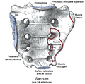
Sacrum.png 680 × 642; 178 KB
-
Sacrum1.png 667 × 663; 276 KB
-
Sarlandière, Jean-Baptiste (Musculi 1).jpg 1,496 × 962; 847 KB
-
Sarlandière, Jean-Baptiste (Musculi 2).jpg 1,495 × 1,038; 878 KB
-
Second Muscular Tabula, Thomas Geminus Wellcome M0012912.jpg 2,614 × 4,331; 2.04 MB
-
Skeletal muscles homo sapiens zh.JPG 1,322 × 2,206; 476 KB
-
Skeletal muscles homo sapiens.JPG 1,650 × 2,206; 422 KB
-
Skeleton and muscles. Wellcome L0010768.jpg 1,442 × 1,288; 502 KB
-
Sketch muscle front and back.jpg 768 × 815; 90 KB
-
Suprahyoideus Muskler.JPG 543 × 657; 54 KB
-
Tertia musculorum tabula (37053621276).jpg 1,896 × 3,346; 1.83 MB
-
The eclectic guide to health; or, Physiology and hygiene (1887) (14761120076).jpg 2,172 × 3,236; 1,016 KB
-
The muscles of the human body, first layer, seen from the ba Wellcome V0007800.jpg 2,302 × 3,200; 3.64 MB
-
The muscles of the human body, second layer, seen from the b Wellcome V0007801.jpg 2,316 × 3,197; 3.35 MB
-
The muscles of the human body, third layer, seen from the ba Wellcome V0007802.jpg 2,311 × 3,195; 3.77 MB
-
The Muscular System from 13th century Wellcome M0017746.jpg 2,770 × 3,997; 3.55 MB
-
Transversal US supraspinatus.jpg 1,552 × 940; 209 KB
-
Transversospinales interspinales enko.svg 300 × 300; 1.11 MB
-
Trapacio-linea-nucal2.jpg 651 × 595; 74 KB
-
Trochlear and frontal nerves.jpg 960 × 720; 100 KB
-
Twelve anatomical figures. Wellcome L0003413.jpg 1,072 × 1,630; 626 KB
-
Two anatomical oil paintings by d'Agoty, 1765-1765 Wellcome V0017123.jpg 2,380 × 3,451; 2.58 MB
-
Two écorchés, facing right. Drawing, attributed to Wellcome V0007723.jpg 2,285 × 3,396; 4.74 MB
-
Two écorchés, one walking with the aid of a staff grasped wi Wellcome V0007953.jpg 3,209 × 2,529; 3.93 MB
-
Vesalius - Muskler.jpg 3,750 × 440; 276 KB
-
Vue antérieure et postérieure des muscles du corps de l'homme.jpg 1,380 × 1,000; 635 KB
-
W. Cowper, Myotomia reformata, 1724 Wellcome L0011884.jpg 1,220 × 1,660; 808 KB
-
W. Cowper, Myotomia reformata; ecorche Wellcome L0024326.jpg 1,142 × 1,680; 836 KB
-
William Orpen, Anatomical Study, Male Torso c.1906. Chalk, Tate.jpg 400 × 561; 49 KB
-
中学校保健 筋肉.png 1,738 × 2,429; 2.58 MB


