Category:Anatomy of the human knee
Jump to navigation
Jump to search
Subcategories
This category has the following 9 subcategories, out of 9 total.
- MRI of the human knee (7 F)
A
B
- Bones of the human knee (28 F)
D
- Dissection of the human knee (22 F)
H
L
M
P
- Popliteal fossa (28 F)
Media in category "Anatomy of the human knee"
The following 92 files are in this category, out of 92 total.
-
3D printed knee 20151126.jpg 1,920 × 2,560; 2.37 MB
-
908 Bursa.jpg 1,360 × 1,091; 671 KB
-
917 Knee Joint.jpg 2,250 × 1,725; 1.11 MB
-
Acute inflammation of the synovial membrane of the knee joint Wellcome L0062592.jpg 4,118 × 4,809; 3.8 MB
-
Anatomía de la rodilla.jpg 1,280 × 800; 367 KB
-
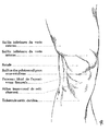
Anterior region of knee. Testut. ro.jpg 499 × 866; 114 KB
-
Articulatio genus - frontal.jpg 4,608 × 3,456; 4.26 MB
-
Articulatio genus - lateral.jpg 4,608 × 3,456; 4.06 MB
-
Articulation du genou.jpg 1,506 × 957; 295 KB
-
Blausen 0596 KneeAnatomy Front Arabic YM.png 1,482 × 1,570; 1.78 MB
-
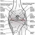
Blausen 0596 KneeAnatomy Front.png 1,500 × 1,500; 1.6 MB
-
Blausen 0597 KneeAnatomy Side-ar.png 1,024 × 768; 672 KB
-
Blausen 0597 KneeAnatomy Side-es.png 1,100 × 825; 1.13 MB
-
Blausen 0597 KneeAnatomy Side-eu.png 450 × 338; 131 KB
-
Blausen 0597 KneeAnatomy Side.png 1,024 × 768; 620 KB
-
Bulletin of the Warren Anatomical Museum (1910) (14763153085).jpg 2,854 × 4,285; 1.42 MB
-
Coupe sagittale du genou (Gray 350).png 1,760 × 2,026; 828 KB
-
Fullsize.jpg 542 × 546; 118 KB
-
Girnelė.png 608 × 456; 1.06 MB
-
Gray345.png 276 × 575; 40 KB
-
Gray346.png 318 × 550; 43 KB
-
Gray347.png 301 × 550; 47 KB
-
Gray348 zh.png 500 × 454; 124 KB
-
Gray348-it.png 1,000 × 1,000; 799 KB
-
Gray348-pa.png 1,000 × 1,000; 898 KB
-
Gray348.png 500 × 454; 42 KB
-
Gray350.png 462 × 600; 49 KB
-
Gray351.png 431 × 550; 39 KB
-
Gray352.png 495 × 500; 43 KB
-
Gray552 es.png 500 × 453; 56 KB
-
Gray552.png 500 × 453; 40 KB
-
Human Knee Anatomy.jpg 960 × 720; 102 KB
-
Human knee joint FE model.png 1,266 × 640; 291 KB
-
Kelio sąnarys.jpg 480 × 360; 6 KB
-
Knee anatomy (Fa).png 800 × 600; 446 KB
-
Knee Biomechanics.JPG 1,303 × 619; 113 KB
-
Knee dorsal.png 427 × 687; 129 KB
-
Knee MRI 113746 rgbcb.png 474 × 498; 342 KB
-
Knee sagittal.png 440 × 784; 205 KB
-
Knee ventral.png 499 × 800; 157 KB
-
Knee XRay.JPG 1,281 × 960; 118 KB
-
KneeElbow (10819138074).jpg 2,025 × 2,963; 1.04 MB
-
KneeHealthyCartilage.jpg 916 × 851; 105 KB
-
KneeHealthyCartilage1.jpg 916 × 851; 83 KB
-
KneeHealthyCartilage2.jpg 916 × 851; 114 KB
-
Kniegelenk, RP-F-F26541.jpg 5,084 × 3,518; 1.51 MB
-
Kniegelenkvorne.png 1,020 × 810; 184 KB
-
Koshba.jpg 300 × 296; 18 KB
-
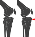
Legamenti crociati.jpg 477 × 574; 109 KB
-
Ligament croisé antérieur.png 333 × 309; 43 KB
-
Ligament croisé postérieur.png 306 × 549; 87 KB
-
Medial Meniscus Injury.png 1,000 × 1,200; 626 KB
-
Medical X-Ray imaging IAD05 nevit.jpg 2,486 × 2,035; 1.77 MB
-
Medical X-Ray imaging NNU06 nevit.jpg 1,784 × 2,384; 414 KB
-
Meniscus of the Knee Unlabeled.jpg 1,000 × 734; 243 KB
-
MRI knee abdonrmal.jpg 523 × 672; 126 KB
-
Obr. anatomie kostí kolenního kloubu.png 1,110 × 832; 420 KB
-
Patella. The Human Project, USA National Library.JPG 764 × 768; 63 KB
-
Plateau du tibia (Gray 349).png 1,618 × 1,166; 536 KB
-
Prepatellar bursa.png 462 × 600; 215 KB
-
Richer - Anatomie artistique, 1 p. 231-1.png 1,540 × 1,824; 38 KB
-
Richer - Anatomie artistique, 1 p. 231-2.png 1,540 × 1,824; 42 KB
-
Richer - Anatomie artistique, 1 p. 232-1.png 1,605 × 1,480; 32 KB
-
Richer - Anatomie artistique, 1 p. 232-2.png 1,605 × 1,480; 32 KB
-
Richer - Anatomie artistique, 1 p. 233.png 996 × 1,588; 21 KB
-
Richer - Anatomie artistique, 1 p. 234.png 2,769 × 1,320; 83 KB
-
Richer - Anatomie artistique, 1 p. 236.png 1,908 × 2,519; 40 KB
-
Richer - Anatomie artistique, 1 p. 237.png 2,117 × 2,893; 44 KB
-
Richer - Anatomie artistique, 1 p. 260.png 3,244 × 1,505; 57 KB
-
-
Slide2umt.JPG 960 × 720; 70 KB
-
Sobo 1909 217.png 1,704 × 2,404; 11.74 MB
-
Sobo 1909 218.png 1,620 × 2,028; 9.42 MB
-
Sobo 1909 219.png 2,028 × 2,512; 14.6 MB
-
Sobo 1909 221.png 1,516 × 984; 1.09 MB
-
Sobo 1909 222.png 1,652 × 1,692; 8.01 MB
-
Staw kolanowy.jpg 259 × 195; 7 KB
-
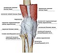
Svyazki-kolennoj-chashechki.jpg 1,582 × 1,443; 446 KB
-
Syndrome rotulien 002.jpg 479 × 420; 20 KB
-
Tape27.png 470 × 500; 67 KB
-
Tape28.png 470 × 500; 59 KB
-
Total knee replacment -lateral X-ray 2008.jpg 2,448 × 3,264; 510 KB
-
Transverse section of knee joint Wellcome L0025028.jpg 1,264 × 1,558; 959 KB
-
Ultrasound Scan ND 084648 0848430 cr.png 457 × 343; 48 KB
-
Ultrasound Scan ND 084648 0850160 cr.png 505 × 379; 75 KB
-
Ultrasound Scan ND 084648 0850340 cr.png 481 × 361; 63 KB
-
Ultrasound Scan ND 084648 0856050 cr.png 643 × 482; 183 KB
-
Virtual palpation.jpg 196 × 240; 30 KB
-
БСЭ1. Коленный сустав.jpg 497 × 395; 145 KB
-
膝の内部構造(右内側).PNG 660 × 800; 104 KB


