Category:Anatomical plates and drawings of the human ulna
Jump to navigation
Jump to search
Media in category "Anatomical plates and drawings of the human ulna"
The following 29 files are in this category, out of 29 total.
-
Bursa of elbow joint.jpg 400 × 550; 76 KB
-
Dorsal tilt.jpg 141 × 181; 5 KB
-
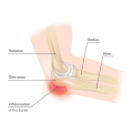
Elbow - Inflammation of the bursa.svg 512 × 512; 176 KB
-
Elbow nerves.svg 512 × 512; 173 KB
-
Elbow subluxation 2.svg 692 × 1,432; 334 KB
-
Elbow subluxation.svg 512 × 512; 330 KB
-
Epicondyluslateralishumeri.png 271 × 600; 251 KB
-
Forearm cut.png 550 × 422; 104 KB
-
Gray212.png 251 × 700; 36 KB
-
Gray213.png 700 × 1,245; 70 KB
-
Gray214.png 717 × 1,254; 93 KB
-
Gray215.png 253 × 450; 9 KB
-
Gray216.png 211 × 500; 20 KB
-
Gray329 numbered.png 400 × 650; 349 KB
-
Gray329.png 313 × 650; 50 KB
-
Gray330.png 277 × 613; 42 KB
-
Gray331.png 252 × 550; 32 KB
-
Gray332-ar.png 249 × 550; 84 KB
-
Gray332.png 249 × 550; 36 KB
-
Gray333.png 404 × 388; 35 KB
-
Gray334.png 509 × 466; 49 KB
-
Gray335.png 508 × 441; 46 KB
-
Gray336.png 550 × 430; 48 KB
-
Ligaments of the elbow.jpg 1,000 × 1,000; 154 KB
-
Olecranon colored.png 251 × 700; 225 KB
-
Radial angulation.jpg 194 × 150; 7 KB
-
Radius and Ulna.jpg 337 × 599; 50 KB
-
Ulna.JPG 416 × 1,726; 104 KB
-
Ulnar variance.jpg 334 × 244; 15 KB


