Category:An introduction to dermatology (1905) book
Jump to navigation
Jump to search
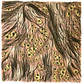
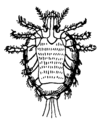
This category is for illustrations from the book: Walker, Norman Purvis (1905) An introduction to dermatology (3rd ed.), William Wood and company Retrieved on 26 September 2010.
which contains 49 plates and 50 illustrations.
Media in category "An introduction to dermatology (1905) book"
The following 101 files are in this category, out of 101 total.
-
An introduction to dermatology (1905) Acitonomycosis.jpg 764 × 1,199; 582 KB
-
An introduction to dermatology (1905) Acne (indurta).jpg 1,232 × 1,928; 978 KB
-
An introduction to dermatology (1905) ALOPECI AREATA.jpg 622 × 626; 257 KB
-
An introduction to dermatology (1905) blastomycosis.jpg 1,296 × 1,592; 741 KB
-
An introduction to dermatology (1905) Bromide Rash.jpg 1,807 × 1,278; 1.01 MB
-
An introduction to dermatology (1905) Bullous eruption Septic Pemphigus.jpg 1,040 × 688; 519 KB
-
An introduction to dermatology (1905) Catarrhal Lupus.jpg 836 × 676; 412 KB
-
An introduction to dermatology (1905) CHEIROPOMPHOLYX.jpg 812 × 1,116; 673 KB
-
An introduction to dermatology (1905) comedo expressor.png 1,760 × 151; 6 KB
-
An introduction to dermatology (1905) Dermatitis herpetiformis.png 1,484 × 1,108; 56 KB
-
An introduction to dermatology (1905) Dermographism.jpg 1,108 × 628; 441 KB
-
An introduction to dermatology (1905) Diagram of Skin.jpg 2,145 × 1,379; 1.59 MB
-
An introduction to dermatology (1905) ecthyma.jpg 1,235 × 1,051; 562 KB
-
An introduction to dermatology (1905) eczema 2.jpg 656 × 1,952; 480 KB
-
An introduction to dermatology (1905) Eczema.png 1,702 × 1,223; 57 KB
-
An introduction to dermatology (1905) Erythema Bullosum.jpg 916 × 1,816; 512 KB
-
An introduction to dermatology (1905) Erythema following vacination.jpg 1,340 × 1,644; 992 KB
-
An introduction to dermatology (1905) erythema induratum 2.jpg 1,086 × 1,542; 473 KB
-
An introduction to dermatology (1905) erythema induratum.jpg 1,050 × 1,507; 582 KB
-
An introduction to dermatology (1905) ERYTHEMA IRIS.jpg 978 × 1,417; 672 KB
-
An introduction to dermatology (1905) ERYTHEMA NODOSUM.jpg 1,172 × 1,920; 701 KB
-
An introduction to dermatology (1905) Favus 2.jpg 1,060 × 1,504; 481 KB
-
An introduction to dermatology (1905) Favus.jpg 627 × 631; 337 KB
-
An introduction to dermatology (1905) Fibroid Lupus.jpg 916 × 584; 453 KB
-
An introduction to dermatology (1905) herpes zoster 2.jpg 902 × 1,193; 371 KB
-
An introduction to dermatology (1905) herpes zoster 3.jpg 880 × 408; 220 KB
-
An introduction to dermatology (1905) herpes zoster.jpg 967 × 1,710; 414 KB
-
An introduction to dermatology (1905) Hydroa gravidarum.jpg 1,199 × 1,790; 1.3 MB
-
An introduction to dermatology (1905) Hydroa vacciniforme.jpg 883 × 1,204; 485 KB
-
An introduction to dermatology (1905) Ichtyosis 2.jpg 1,280 × 424; 286 KB
-
An introduction to dermatology (1905) Ichtyosis 3.jpg 1,292 × 1,016; 543 KB
-
An introduction to dermatology (1905) Ichtyosis diagram.jpg 640 × 512; 218 KB
-
An introduction to dermatology (1905) Ichtyosis.jpg 1,252 × 1,868; 1,010 KB
-
An introduction to dermatology (1905) Impetigo circinata.jpg 801 × 1,103; 435 KB
-
An introduction to dermatology (1905) Impetigo contagioso.jpg 1,268 × 1,992; 1.12 MB
-
An introduction to dermatology (1905) keloid.jpg 1,024 × 1,428; 929 KB
-
An introduction to dermatology (1905) kerion.jpg 1,062 × 799; 452 KB
-
An introduction to dermatology (1905) Lichen planus 1.jpg 1,180 × 1,808; 576 KB
-
An introduction to dermatology (1905) Lichen planus 2.jpg 932 × 1,788; 418 KB
-
An introduction to dermatology (1905) lupus erythematosus 2.jpg 1,056 × 1,780; 789 KB
-
An introduction to dermatology (1905) Lupus erythematosus section.jpg 696 × 568; 283 KB
-
An introduction to dermatology (1905) lupus erythematosus.jpg 688 × 612; 281 KB
-
An introduction to dermatology (1905) Lupus vulgaris 2.jpg 1,312 × 1,916; 1 MB
-
An introduction to dermatology (1905) Lupus vulgaris simplex.jpg 900 × 644; 419 KB
-
An introduction to dermatology (1905) maculo-anaesthetic leprosy.jpg 894 × 1,839; 639 KB
-
An introduction to dermatology (1905) Miliaria 80x.png 1,440 × 840; 33 KB
-
An introduction to dermatology (1905) Molluscum Contagiosum section.png 1,404 × 1,212; 86 KB
-
An introduction to dermatology (1905) molluscum fibrosum 1.jpg 917 × 1,525; 711 KB
-
An introduction to dermatology (1905) Mycosis Fungoides later stage.jpg 1,486 × 1,039; 484 KB
-
An introduction to dermatology (1905) Mycosis Fungoides.jpg 1,054 × 1,398; 849 KB
-
An introduction to dermatology (1905) nodular leprosy.jpg 1,324 × 1,488; 1.28 MB
-
An introduction to dermatology (1905) Part of a hair affected by Favus.png 1,963 × 396; 19 KB
-
An introduction to dermatology (1905) pediculus capitis 42x.jpg 216 × 428; 68 KB
-
An introduction to dermatology (1905) Pediculus corporis 50x.jpg 428 × 216; 66 KB
-
An introduction to dermatology (1905) Pediculus pubis 50x.png 864 × 984; 16 KB
-
An introduction to dermatology (1905) Pemphigus.jpg 1,211 × 896; 604 KB
-
An introduction to dermatology (1905) pityriasis rosea diagram.png 1,305 × 744; 27 KB
-
An introduction to dermatology (1905) Pityriasis rosea.jpg 1,212 × 1,732; 743 KB
-
An introduction to dermatology (1905) Primula obconica(P. poculifornis).jpg 924 × 1,372; 794 KB
-
An introduction to dermatology (1905) Psoriasis (partly treated).jpg 1,108 × 1,808; 697 KB
-
An introduction to dermatology (1905) Psoriasis.jpg 1,159 × 1,636; 610 KB
-
An introduction to dermatology (1905) Rhus toxicodendron.jpg 1,020 × 1,348; 638 KB
-
An introduction to dermatology (1905) ringworm photo.jpg 844 × 565; 93 KB
-
An introduction to dermatology (1905) Ringworm.jpg 627 × 851; 259 KB
-
An introduction to dermatology (1905) rodent ulcer.jpg 1,220 × 1,760; 1.21 MB
-
An introduction to dermatology (1905) scabies.jpg 1,028 × 1,296; 471 KB
-
An introduction to dermatology (1905) scleroderma.jpg 1,424 × 1,956; 854 KB
-
An introduction to dermatology (1905) Seborrhcea 2.jpg 1,172 × 2,016; 1.11 MB
-
An introduction to dermatology (1905) Seborrhcea.jpg 1,464 × 1,961; 1.38 MB
-
An introduction to dermatology (1905) Section from rodent ulcer.jpg 896 × 564; 426 KB
-
An introduction to dermatology (1905) Section of a scutulum in situ.jpg 816 × 396; 236 KB
-
An introduction to dermatology (1905) section of early lesion.png 1,568 × 912; 38 KB
-
An introduction to dermatology (1905) syphilis (secondary).jpg 1,312 × 1,896; 789 KB
-
An introduction to dermatology (1905) syphilis (tertiary).jpg 880 × 1,920; 770 KB
-
An introduction to dermatology (1905) transverse section of nail.png 1,928 × 804; 52 KB
-
An introduction to dermatology (1905) tuberculosis fig. 45.jpg 1,153 × 746; 493 KB
-
An introduction to dermatology (1905) Vessicle in the prickle layer.jpg 636 × 460; 242 KB
-
An introduction to dermatology (1905) vitiligo or leucoderma.jpg 874 × 1,733; 320 KB
-
An introduction to dermatology (1905) xanthoma diabetacorum.jpg 380 × 1,848; 260 KB


