File:Pseudorhabdosynochus caledonicus body.jpg
Jump to navigation
Jump to search

Size of this preview: 378 × 599 pixels. Other resolutions: 151 × 240 pixels | 303 × 480 pixels | 900 × 1,425 pixels.
Original file (900 × 1,425 pixels, file size: 359 KB, MIME type: image/jpeg)
Captions
Captions
Add a one-line explanation of what this file represents
Summary
[edit]
| Description |
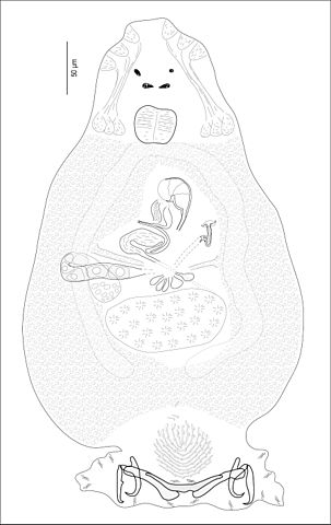
English: Pseudorhabdosynochus caledonicus Justine, 2005 (Monogenea, Diplectanidae) a parasite of the grouper Epinephelus fasciatus (Perciformes, Serranidae) off New Caledonia. Holotype, whole body, ventral view. Original line drawing. Species described in article: Justine, 2005, Systematic Parasitology 62, 1-37. Preview
See Pseudorhabdosynochus caledonicus male and female organs.jpg for sclerotised male and female copulatory organs of same species. Français : Pseudorhabdosynochus caledonicus Justine, 2005 (Monogenea, Diplectanidae) parasite du mérou Epinephelus fasciatus (Perciformes, Serranidae) au large de la Nouvelle-Calédonie. Holotype, corps entier, vue ventrale. Dessin au trait original. Espèce décrite dans l'article: Justine, 2005, Systematic Parasitology 62, 1-37. Preview
Voir Pseudorhabdosynochus caledonicus male and female organs.jpg pour les organes copulateurs sclérifiés de la même espèce. |
| Date | |
| Source | Own work |
| Author | Jean-Lou Justine |
Licensing
[edit]
I, the copyright holder of this work, hereby publish it under the following license:
This file is licensed under the Creative Commons Attribution-Share Alike 3.0 Unported license.
- You are free:
- to share – to copy, distribute and transmit the work
- to remix – to adapt the work
- Under the following conditions:
- attribution – You must give appropriate credit, provide a link to the license, and indicate if changes were made. You may do so in any reasonable manner, but not in any way that suggests the licensor endorses you or your use.
- share alike – If you remix, transform, or build upon the material, you must distribute your contributions under the same or compatible license as the original.
| Annotations | This image is annotated: View the annotations at Commons |
File history
Click on a date/time to view the file as it appeared at that time.
| Date/Time | Thumbnail | Dimensions | User | Comment | |
|---|---|---|---|---|---|
| current | 19:58, 2 April 2012 | | 900 × 1,425 (359 KB) | Jeanloujustine (talk | contribs) |
You cannot overwrite this file.
File usage on Commons
The following 2 pages use this file:
File usage on other wikis
The following other wikis use this file:
- Usage on ceb.wikipedia.org
- Usage on en.wikipedia.org
- Usage on nl.wikipedia.org
- Usage on species.wikimedia.org
- Usage on www.wikidata.org