File:Mouse embryonic salivary gland growing in vitro.jpg
Mouse_embryonic_salivary_gland_growing_in_vitro.jpg (548 × 549 pixels, file size: 169 KB, MIME type: image/jpeg)
Captions
Captions
Summary
[edit]| DescriptionMouse embryonic salivary gland growing in vitro.jpg |
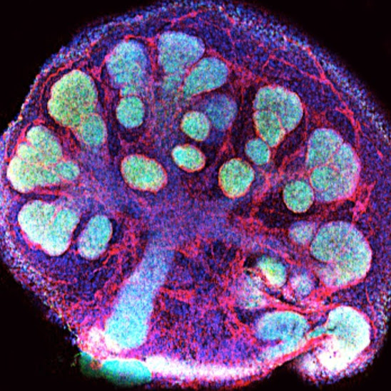
English: This is an example of growing a mouse embryonic organ in vitro in order to understand how they grow and how to regenerate organs. This is an embryonic mouse salivary gland that develops via branching morphogenesis, and makes ducts and buds that will give rise to the functional organ (a salivary gland). Green: marker for epithelial cells (E-Cadherin); Red: marker for matrix component that cells excrete to attach to each other (Collagen-IV); Blue: marker for nuclei of the cells (DNA marker ToPro3). This is a confocal microscopy (stacked collection of several microscopic photos). We can grow organs in vitro for studying regenerative biomedical strategies in the future. English: This is an example of growing mouse embryonic organs in vitro in order to understand how they grow and how to regenerate organs. This is an embryonic mouse salivary gland that develops via branching morphogenesis, and makes ducts and buds that will give rise to the functional organ (a salivary gland). Green: marker for epithelial cells (E-Cadherin); Red: marker for matrix component that cells excrete to attach to each other (Collagen-IV); Blue: marker for nuclei of the cells (DNA marker ToPro3). This is a confocal microscopy (stacked collection of several microscopic photos). We can grow organs in vitro for studying regenerative biomedical strategies in the future. |
| Date | |
| Source | Own work |
| Author | Irebustini |
This picture was taken in a research laboratory in the National Institute of Dental and Craniofacial Research in Bethesda, MD.
Licensing
[edit]- You are free:
- to share – to copy, distribute and transmit the work
- to remix – to adapt the work
- Under the following conditions:
- attribution – You must give appropriate credit, provide a link to the license, and indicate if changes were made. You may do so in any reasonable manner, but not in any way that suggests the licensor endorses you or your use.
| This image was uploaded as part of Wiki Science Competition 2017. |
File history
Click on a date/time to view the file as it appeared at that time.
| Date/Time | Thumbnail | Dimensions | User | Comment | |
|---|---|---|---|---|---|
| current | 15:03, 3 November 2017 |  | 548 × 549 (169 KB) | Irebustini (talk | contribs) | User created page with UploadWizard |
You cannot overwrite this file.
File usage on Commons
The following 24 pages use this file:
- Commons:Wiki Science Competition 2017
- Commons:Wiki Science Competition 2017/Images
- Commons:Wiki Science Competition 2017/Images/ar
- Commons:Wiki Science Competition 2017/Images/da
- Commons:Wiki Science Competition 2017/Images/de
- Commons:Wiki Science Competition 2017/Images/en
- Commons:Wiki Science Competition 2017/Images/fr
- Commons:Wiki Science Competition 2017/Images/it
- Commons:Wiki Science Competition 2017/Images/mk
- Commons:Wiki Science Competition 2017/Images/pl
- Commons:Wiki Science Competition 2017/ar
- Commons:Wiki Science Competition 2017/da
- Commons:Wiki Science Competition 2017/de
- Commons:Wiki Science Competition 2017/en
- Commons:Wiki Science Competition 2017/es
- Commons:Wiki Science Competition 2017/fr
- Commons:Wiki Science Competition 2017/it
- Commons:Wiki Science Competition 2017/mk
- Commons:Wiki Science Competition 2017/pl
- Commons:Wiki Science Competition 2017/pt-br
- Commons:Wiki Science Competition 2017/ru
- Commons:Wiki Science Competition 2017/th
- Commons:Wiki Science Competition 2017/tr
- Commons:Wiki Science Competition 2017/vi
File usage on other wikis
The following other wikis use this file:
- Usage on uk.wikipedia.org
Metadata
This file contains additional information such as Exif metadata which may have been added by the digital camera, scanner, or software program used to create or digitize it. If the file has been modified from its original state, some details such as the timestamp may not fully reflect those of the original file. The timestamp is only as accurate as the clock in the camera, and it may be completely wrong.
| Width | 1,129 px |
|---|---|
| Height | 1,157 px |
| Bits per component |
|
| Pixel composition | RGB |
| Orientation | Normal |
| Number of components | 3 |
| Horizontal resolution | 300 dpi |
| Vertical resolution | 300 dpi |
| Software used | Adobe Photoshop CS5.1 Macintosh |
| File change date and time | 16:54, 29 February 2012 |
| Exif version | 2.21 |
| Color space | sRGB |
| Unique image ID | 74450d0f026b99770000000000000000 |
| Date and time of digitizing | 04:55, 23 February 2012 |
| Date metadata was last modified | 11:54, 29 February 2012 |
| Unique ID of original document | xmp.did:05801174072068118A6DDE39EE8383E3 |

