File:Hepatitis B virus v2 (2).svg

Original file (SVG file, nominally 754 × 428 pixels, file size: 81 KB)
Captions
Captions
Contents
Summary
[edit]| DescriptionHepatitis B virus v2 (2).svg |
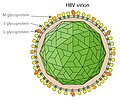
English: Simplified drawing of the Hepatitis B virus particle and surface (surplus) antigen. Created by en:User:GrahamColm |
| Date | 14 November 2007 (original upload date) |
| Source | Transferred from en.wikipedia |
| Author | Original uploader was TimVickers at en.wikipedia |
| Permission (Reusing this file) |
Released into the public domain (by the author). |
| Other versions |
[edit] |
| SVG development InfoField | The source code of this SVG is uncheckable. This W3C-uncheckable vector image was created with another SVG tool. WMF tells W3 the SVG file is image/png! |
Extended description
[edit]The structure of the Hepatitis B virus as first described by Dane & al.[1] and Jokelainen, Krohn & al.[2] in 1970:
Virion
[edit]The hepatitis B virion, is a complex, spherical, double shelled particle with a diameter of 42 nm.[1][2][3]
- The 6 nm[2] thick outer viral envelope or membrane contains host-derived lipids and surface proteins,[2] known collectively as HBsAg.[3] The membrane contains globular subunits each measuring ca. 3 to 4 nm in diameter and 3 to 4 nm apart.[2]
- Within the membrane sphere is a 2 nm thick icosahedral nucleocapsid inner core composed of protein (HBcAg) with a diameter of 27 nm.[2] When viewed through an electron microscope the inner core may appear pentagonal or hexagonal,[2] depending on the relative position of the sample.
- The nucleocapsid contains a viral genome[2] of circular, partially double stranded DNA[3] and endogenous DNA polymerase[4][3] within a diameter of ca. 18 nm.[2]
The virion was initially referred to as the Dane particle.[4] Only after Baruch Blumberg received the Nobel Prize in Medicine in 1976 was it universally accepted that the particle is a virus and the infectious agent of Hepatitis B.
Australia antigen (HBsAg)
[edit]The serum of infected patients also contain small spherical and rod-shaped particles with a diameter of ca. 20 nm,[5] consisting of surplus virus-coat material containing the HBsAg antigen.[1][2] This antigen was first discovered by Baruch Blumberg in 1965 in the blood of Australian aboriginal people and initially known as "Australia antigen".[6] It was shown to be associated with "serum hepatitis" by A. M. Prince in 1968.[7]
The outer membrane of the virion is sometimes extended as a tubular tail on one side of the virus particle (not shown);[2][3] these virion "tails" are identical to the small particles.[2][3]
The hepatitis B e antigens (shown) are considered not part of the viral particle.
References
[edit]- ↑ a b c D.S. Dane , C.H. Cameron , Moya Briggs (1970). "Virus-Like Particles in Serum of Patients with Australia-Antigen-Associated Hepatitis". The Lancet 295: 695–698. DOI:10.1016/S0140-6736(70)90926-8.
- ↑ a b c d e f g h i j k l P. T. Jokelainen, Kai Krohn, A. M. Prince and N. D. C. Finlayson (1970). "Electron Microscopic Observations on Virus-Like Particles Associated with SH Antigen". J Virol. 6 (5): 685-689. ISSN 1098-5514.
- ↑ a b c d e f The hepatitis B virus. WHO.
- ↑ a b Almeida J D, Rubenstein D & Scott E J. (1971). "New antigen-antibody system in Australia-antigen-positive hepatitis". The Lancet 298 (7736): 1225–7. DOI:10.1016/S0140-6736(71)90543-5.
- ↑ Bayer, M. E., B. S. Blumberg, and B. Werner (1968). "Particles associated with Australia antigen in the sera of patients with leukemia, Down's syndrome and hepatitis.". Nature (London) 218: 1057-1059.
- ↑ Baruch S. Blumberg, Harvey J. Alter, and Sam Visnich (Jul 1984). "Landmark article Feb 15, 1965: A 'new' antigen in leukemia sera. By Baruch S. Blumberg, Harvey J. Alter, and Sam Visnich". JAMA 252 (2): 252–7. DOI:10.1001/jama.252.2.252. PMID 6374187. ISSN 0098-7484.
- ↑ Prince, A. M. (1968). "An antigen detected in the blood during the incubation period of serum hepatitis". Proc. Nat. Acad. Sci. U.S.A. 60: 814-821.
Licensing
[edit]| Public domainPublic domainfalsefalse |
| |
This work has been released into the public domain by its author, TimVickers, at the English Wikipedia project. This applies worldwide. In case this is not legally possible: |
Original upload log
[edit]- 2007-11-14 18:14 TimVickers 843×577× (81917 bytes) Simplified drawing of the Hepatitis B virus particle and surface (surplus) antigen
File history
Click on a date/time to view the file as it appeared at that time.
| Date/Time | Thumbnail | Dimensions | User | Comment | |
|---|---|---|---|---|---|
| current | 18:36, 23 January 2013 |  | 754 × 428 (81 KB) | Graham Beards (talk | contribs) | Reverted to version as of 02:06, 10 December 2009 |
| 02:06, 10 December 2009 |  | 754 × 428 (81 KB) | Huckfinne (talk | contribs) | Made it into a .svg file and improved the labeling to the standards I've learned in medical school. |
You cannot overwrite this file.
File usage on Commons
The following 8 pages use this file:
File usage on other wikis
The following other wikis use this file:
- Usage on en.wikipedia.org
- Usage on ha.wikipedia.org
Metadata
This file contains additional information such as Exif metadata which may have been added by the digital camera, scanner, or software program used to create or digitize it. If the file has been modified from its original state, some details such as the timestamp may not fully reflect those of the original file. The timestamp is only as accurate as the clock in the camera, and it may be completely wrong.
| Width | 754.23553 |
|---|---|
| Height | 428.01526 |


