File:Hemoglobin F.png
From Wikimedia Commons, the free media repository
Jump to navigation
Jump to search
Hemoglobin_F.png (600 × 600 pixels, file size: 294 KB, MIME type: image/png)
File information
Structured data
Captions
Captions
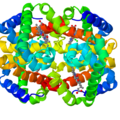
Fetal hemoglobin (HbF), formed by two alpha subunits (top) and two gamma subunits (bottom). The 4 heme groups are also displayed. All chains (ribbons) are rainbow-colored from blue to red (N- to C-termini)
Структура фетального гемоглобинa (HbF), образованный двумя альфа-субъединицами (вверху) и двумя гамма-субъединицами (внизу). Также показаны 4 гемовые группы. Все цепи (ленты) окрашены в цвета радуги от синего до красного (от N- до C-концов)
| This image appeared on English Wikipedia's Main Page in the Did you know? column on 13 April 2004 (see archives). |
Summary
[edit]| DescriptionHemoglobin F.png |
English: Fetal hemoglobin (HbF), formed by two alpha subunits (top) and two gamma subunits (bottom). The 4 heme groups are also displayed. All chains (ribbons) are rainbow-colored from blue to red (N- to C-termini) |
| Date | 10 February 2016 (original upload date) |
| Source | Own work - A rendering prepared by me, using Jmol, from 4MQJ at PDB (doi:10.2210/pdb4mqj/pdb). |
| Author | AngelHerraez |
Licensing
[edit]| Public domainPublic domainfalsefalse |
| I, the copyright holder of this work, release this work into the public domain. This applies worldwide. In some countries this may not be legally possible; if so: I grant anyone the right to use this work for any purpose, without any conditions, unless such conditions are required by law. |
Original upload log
[edit]The original description page was here. All following user names refer to en.wikipedia.
- 2004-04-12 15:39 Diberri 612×522× (25345 bytes) Fetal hemoglobin (transparent background). My snapshot of rendered version from PDB file [http://www.snv.jussieu.fr/vie/telecharge/pdb/HbF-1FDH.pdb].
- 2004-04-12 15:01 Diberri 612×522× (36662 bytes) Fetal hemoglobin (white background). My snapshot of rendered version from PDB file [http://www.snv.jussieu.fr/vie/telecharge/pdb/HbF-1FDH.pdb].
File history
Click on a date/time to view the file as it appeared at that time.
| Date/Time | Thumbnail | Dimensions | User | Comment | |
|---|---|---|---|---|---|
| current | 12:25, 10 February 2016 |  | 600 × 600 (294 KB) | AngelHerraez (talk | contribs) | the former image was partially clipped |
| 12:21, 10 February 2016 |  | 600 × 600 (360 KB) | AngelHerraez (talk | contribs) | The prevoius image was a dimer (alpha, gamma) and hence may be misleading. The actual, biologically relevant, structure of hemoglobin F (fetal) is a tetramer (2 alpha + 2 gamma subunits). This rendering displays the tetramer, as well as the 4 heme grou... | |
| 07:06, 16 January 2012 |  | 612 × 522 (25 KB) | File Upload Bot (Magnus Manske) (talk | contribs) | {{BotMoveToCommons|en.wikipedia|year={{subst:CURRENTYEAR}}|month={{subst:CURRENTMONTHNAME}}|day={{subst:CURRENTDAY}}}} {{Information |Description={{en|Fetal hemoglobin (white background).}} |Source=Transferred from [http://en.wikipedia.org en.wikipedia]; |
You cannot overwrite this file.
File usage on Commons
There are no pages that use this file.
File usage on other wikis
The following other wikis use this file:
- Usage on ar.wikipedia.org
- Usage on ca.wikipedia.org
- Usage on en.wikipedia.org
- Usage on en.wikibooks.org
- Usage on fr.wikipedia.org
- Usage on it.wikipedia.org
- Usage on ru.wikipedia.org
- Usage on sh.wikipedia.org
- Usage on sr.wikipedia.org
- Usage on uk.wikipedia.org
Metadata
This file contains additional information such as Exif metadata which may have been added by the digital camera, scanner, or software program used to create or digitize it. If the file has been modified from its original state, some details such as the timestamp may not fully reflect those of the original file. The timestamp is only as accurate as the clock in the camera, and it may be completely wrong.
| Software used | |
|---|---|
| Date and time of digitizing |
|
Structured data
Items portrayed in this file
depicts
some value
10 February 2016
301,544 byte
600 pixel
600 pixel
image/png
1ba491b9a55606ad20d91578cecea55292e76022
Hidden categories:

