File:Elife-25940-fig8-v2.jpg
Jump to navigation
Jump to search

Size of this preview: 620 × 600 pixels. Other resolutions: 248 × 240 pixels | 496 × 480 pixels | 794 × 768 pixels | 1,059 × 1,024 pixels | 1,882 × 1,820 pixels.
Original file (1,882 × 1,820 pixels, file size: 500 KB, MIME type: image/jpeg)
Captions
Captions
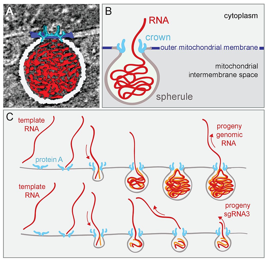
Model of Flock house nodavirus (FHV) replication complex structure and function.
Summary
[edit]
| Description |
English: Model of Flock house nodavirus (FHV) replication complex structure and function.
(A) Amira-assisted 3D segmentation of a single virus replication compartment in FHV-infected cells. The spherule membrane (white) encloses the viral RNA (red) and is anchored to the mitochondrial outer membrane (dark blue), by the FHV protein A crown (light blue, derived from the subtomogram average). (B) Cartoon rendering of the image in (A). Viral RNA is shown as exiting the spherule to include the observation of fibrillary extrusions in many other tomograms. (C) Model of the FHV RNA complex and RNA synthesis, where positive-strand RNA (red) associates with protein A on the mitochondrial membrane to initiate the synthesis of negative-strand RNA (orange) and progeny positive-strand RNA accommodated by increasing volume of the spherule as if blowing up a balloon. In this model, subgenomic RNA3 results from attenuated negative strand synthesis and subse- quent positive-strand production in small spherules. |
| Date | |
| Source |
eLife 2017;6:e25940 DOI: 10.7554/eLife.25940 https://elifesciences.org/articles/25940 |
| Author | Kenneth J Ertel, Desirée Benefield, Daniel Castaño-Diez, Janice G Pennington, Mark Horswill, Johan A den Boon, Marisa S Otegui, and Paul Ahlquist |
Licensing
[edit]
This file is licensed under the Creative Commons Attribution 4.0 International license.
- You are free:
- to share – to copy, distribute and transmit the work
- to remix – to adapt the work
- Under the following conditions:
- attribution – You must give appropriate credit, provide a link to the license, and indicate if changes were made. You may do so in any reasonable manner, but not in any way that suggests the licensor endorses you or your use.
File history
Click on a date/time to view the file as it appeared at that time.
| Date/Time | Thumbnail | Dimensions | User | Comment | |
|---|---|---|---|---|---|
| current | 10:13, 2 December 2020 | | 1,882 × 1,820 (500 KB) | Guest2625 (talk | contribs) | Uploaded a work by Kenneth J Ertel, Desirée Benefield, Daniel Castaño-Diez, Janice G Pennington, Mark Horswill, Johan A den Boon, Marisa S Otegui, and Paul Ahlquist from eLife 2017;6:e25940 DOI: 10.7554/eLife.25940 https://elifesciences.org/articles/25940 with UploadWizard |
You cannot overwrite this file.
File usage on Commons
There are no pages that use this file.