File:Brodmann areas in coronal sections of monkey (Cebus).jpg
Jump to navigation
Jump to search

Size of this preview: 450 × 599 pixels. Other resolutions: 180 × 240 pixels | 360 × 480 pixels | 577 × 768 pixels | 1,200 × 1,598 pixels.
Original file (1,200 × 1,598 pixels, file size: 198 KB, MIME type: image/jpeg)
Captions
Captions
Add a one-line explanation of what this file represents
| Description |
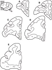
English: Coronal sections showing areal borders. Drawings of coronal sections through five different rostrocaudal levels of Cebus left hemisphere, showing areal borders. Brodmann areas in prefrontal cortex of Cebus depicted. 日本語: オマキザル属のサルの脳の解剖。前頭皮質の冠状断面におけるブロードマン領野。 |
||
| Date | |||
| Source | Roelf J Cruz-Rizzolo, Miguel AX De Lima, Edilson Ervolino, José A de Oliveira and Claudio A Casatti (2011) Cyto-, myelo- and chemoarchitecture of the prefrontal cortex of the Cebus monkey BMC Neuroscience 2011, 12:6 doi:10.1186/1471-2202-12-6 | ||
| Author | Roelf J Cruz-Rizzolo, Miguel AX De Lima, Edilson Ervolino, José A de Oliveira and Claudio A Casatti (2011) | ||
| Permission (Reusing this file) |
|
||
| Other versions |
.png) |
File history
Click on a date/time to view the file as it appeared at that time.
| Date/Time | Thumbnail | Dimensions | User | Comment | |
|---|---|---|---|---|---|
| current | 15:10, 17 August 2012 | | 1,200 × 1,598 (198 KB) | Was a bee (talk | contribs) | {{Information |Description={{en|Coronal sections showing areal borders. Drawings of coronal sections through five different rostrocaudal levels of Cebus left hemisphere, showing areal borders. Brodmann areas in prefrontal cortex of ''Cebus'' depict... |
You cannot overwrite this file.
File usage on Commons
The following page uses this file:
.jpg&oldid=834805684)