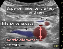
File:Axial plane ultrasound at the navel.jpg
Axial_plane_ultrasound_at_the_navel.jpg (618 × 486 pixels, file size: 77 KB, MIME type: image/jpeg)
Captions
Captions
Summary
[edit]| DescriptionAxial plane ultrasound at the navel.jpg |
English: Abdominal ultrasound in the axial plane at the level of the navel of a 31 year old man, showing normal anatomy. It uses compression to decrease the distance to the aorta for better visualization, resulting in flat shapes of the inferior mesenteric vein and the inferior vena cava. |
| Date | |
| Source | Own work |
| Author |
 - Reusing images - Conflicts of interest: None Consent note: Written informed consent was obtained from the individual, including online publication. |
| Other versions |
 |
Licensing
[edit]| This file is made available under the Creative Commons CC0 1.0 Universal Public Domain Dedication. | |
| The person who associated a work with this deed has dedicated the work to the public domain by waiving all of their rights to the work worldwide under copyright law, including all related and neighboring rights, to the extent allowed by law. You can copy, modify, distribute and perform the work, even for commercial purposes, all without asking permission.
http://creativecommons.org/publicdomain/zero/1.0/deed.enCC0Creative Commons Zero, Public Domain Dedicationfalsefalse |
File history
Click on a date/time to view the file as it appeared at that time.
| Date/Time | Thumbnail | Dimensions | User | Comment | |
|---|---|---|---|---|---|
| current | 19:34, 24 January 2018 |  | 618 × 486 (77 KB) | Mikael Häggström (talk | contribs) | User created page with UploadWizard |
You cannot overwrite this file.
File usage on Commons
The following page uses this file:
Metadata
This file contains additional information such as Exif metadata which may have been added by the digital camera, scanner, or software program used to create or digitize it. If the file has been modified from its original state, some details such as the timestamp may not fully reflect those of the original file. The timestamp is only as accurate as the clock in the camera, and it may be completely wrong.
| JPEG file comment | Intel(R) IPP JPEG encoder [7.0.998] - Sep 2 2010 |
|---|---|
| Orientation | Normal |
