File:3D-SIM-1 NPC Confocal vs 3D-SIM.jpg

Original file (1,069 × 848 pixels, file size: 307 KB, MIME type: image/jpeg)
Captions
Captions
Summary
[edit]| Description3D-SIM-1 NPC Confocal vs 3D-SIM.jpg |
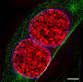
English: Comparison of resolution obtained by confocal laser scanning microscopy (clsm, left) and 3D structured illumination microscopy (3D-SIM-Microscopy, right). Top images each show a nucleus of a mouse cell, the white boxes are magnified at the bottom. Nuclear pores (anti-NPC, red) nuclear envelope (anti-Lamin, green). Chromatin/DNA (DAPI, blue). scale bars: top 5 µm, bottom 1µm.
For further information see: Schermelleh L, Carlton PM, Haase S, Shao L, Winoto L, Kner P, Burke B, Cardoso MC, Agard DA, Gustafsson MG, Leonhardt H, Sedat JW (June 2008). "Subdiffraction multicolor imaging of the nuclear periphery with 3D structured illumination microscopy". Science (journal) 320 (5881): 1332–6. DOI:10.1126/science.1156947. PMID 18535242.
Deutsch: Vergleich des Auflösungsvermögen von konfokaler Laser-Scanning Mikroskopie (CLSM, links) und 3D-SIM (rechts). Zellkernporen (anti-NPC, rot), Zellkernhülle (anti-Lamin B, grün), sowie DNA verpackt in Chromatin (DAPI, blau) wurden in einer Mauszelle simultan angefärbt. Der Maßstab entspricht 5 µm (oben) und 1 µm (unten). Für weitere Informationen siehe oben zitierte Veröffentlichung.) |
||
| Date | |||
| Source | Lothar Schermelleh | ||
| Author | Lothar Schermelleh | ||
| Permission (Reusing this file) |
This file is licensed under the Creative Commons Attribution-Share Alike 3.0 Unported license.
|
||
| Other versions |
 |
3D-SIM images
[edit]| Annotations InfoField | This image is annotated: View the annotations at Commons |
scale bar: 5 µm
File history
Click on a date/time to view the file as it appeared at that time.
| Date/Time | Thumbnail | Dimensions | User | Comment | |
|---|---|---|---|---|---|
| current | 17:21, 16 January 2009 |  | 1,069 × 848 (307 KB) | Dietzel65 (talk | contribs) | {{Information |Description={{en|1=(to be added soon)}} {{de|1=Vergleich für das Auflösungsvermögen von konfokaler Laser-Scanning Mikroskopie (CLSM, links) und 3D-SIM (rechts). Zellkernporen (NPC, rot), Zellkernhülle (Lamin B, grün), sowie DNA |
You cannot overwrite this file.
File usage on Commons
The following 6 pages use this file:
File usage on other wikis
The following other wikis use this file:
- Usage on de.wikipedia.org
- Usage on en.wikipedia.org
- Usage on nl.wikibooks.org
- Usage on uk.wikipedia.org
- Usage on vi.wikipedia.org
- Usage on zh.wikipedia.org
Metadata
This file contains additional information such as Exif metadata which may have been added by the digital camera, scanner, or software program used to create or digitize it. If the file has been modified from its original state, some details such as the timestamp may not fully reflect those of the original file. The timestamp is only as accurate as the clock in the camera, and it may be completely wrong.
| Orientation | Normal |
|---|---|
| Horizontal resolution | 300 dpi |
| Vertical resolution | 300 dpi |
| Software used | Adobe Photoshop CS3 Windows |
| File change date and time | 10:45, 15 January 2009 |
| Color space | sRGB |
| Image width | 1,069 px |
| Image height | 848 px |
| Date and time of digitizing | 11:45, 15 January 2009 |
| Date metadata was last modified | 11:45, 15 January 2009 |
| IIM version | 111 |


