Category:Mikael Häggström/Macropathology
Jump to navigation
Jump to search
These images were created by Mikael Häggström, M.D.
- User info
- Reusing images
Subcategories
This category has the following 2 subcategories, out of 2 total.
Media in category "Mikael Häggström/Macropathology"
The following 75 files are in this category, out of 75 total.
-
Adrenal glands.jpg 2,225 × 957; 566 KB
-
Arachnoid granulations.jpg 3,137 × 2,473; 1.43 MB
-
Arterial aneurysm.jpg 1,009 × 1,093; 401 KB
-
Brain with arachnoid and pia mater.jpg 1,720 × 1,673; 695 KB
-
Copper IUD.jpg 1,905 × 3,537; 1.6 MB
-
Excised melanoma in situ.jpg 408 × 247; 24 KB
-
Gross pathology of a corpus luteum cyst with bleeding.jpg 2,518 × 2,006; 1.18 MB
-
Gross pathology of a mesenteric lymph node.jpg 1,249 × 1,401; 552 KB
-
Gross pathology of a one week old myocardial infarction with focal rupture.jpg 4,437 × 1,589; 1.97 MB
-
Gross pathology of a peritoneal loose body.jpg 1,345 × 1,005; 232 KB
-
Gross pathology of a postmortem blood clot.jpg 1,569 × 1,257; 388 KB
-
Gross pathology of a trabeculated gallbladder, with enlarged inset.jpg 1,737 × 874; 479 KB
-
Gross pathology of a trabeculated gallbladder.jpg 4,032 × 3,024; 1.19 MB
-
Gross pathology of a uterus with extensive adenomyosis.jpg 1,082 × 581; 181 KB
-
Gross pathology of amputated finger after longitudinal cutting.jpg 2,409 × 2,109; 1.46 MB
-
Gross pathology of amputated finger sections in cassette.jpg 2,225 × 1,277; 831 KB
-
Gross pathology of an accessory spleen.jpg 3,852 × 2,597; 1.92 MB
-
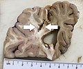
Gross pathology of an old cerebral stroke, annotated.jpg 3,117 × 2,601; 1.79 MB
-
Gross pathology of an old cerebral stroke.jpg 3,117 × 2,601; 1.8 MB
-
Gross pathology of appendicitis containing a blood-tinged purulent exudate.jpg 2,817 × 2,017; 1.17 MB
-
Gross pathology of appendicitis with a patchy purulent exudate, annotated.jpg 1,193 × 945; 329 KB
-
Gross pathology of appendicitis with a patchy purulent exudate.jpg 2,567 × 2,202; 1.41 MB
-
Gross pathology of brain after sectioning.jpg 2,869 × 2,117; 1.68 MB
-
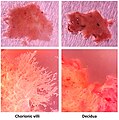
Gross pathology of chorionic villi and decidua.jpg 3,497 × 3,521; 2.81 MB
-
Gross pathology of chorionic villi at 10 weeks.jpg 1,075 × 897; 311 KB
-
Gross pathology of chorionic villi.jpg 697 × 643; 92 KB
-
Gross pathology of diverticulitis with diverticular abscesses.jpg 2,201 × 1,721; 1.19 MB
-
Gross pathology of diverticulosis.jpg 2,517 × 1,597; 852 KB
-
Gross pathology of endometrial adenocarcinoma.jpg 2,825 × 1,406; 1.35 MB
-
Gross pathology of fetal membranes versus decidua.jpg 1,823 × 805; 417 KB
-
Gross pathology of fixed chorionic villi.jpg 899 × 717; 93 KB
-
Gross pathology of fresh chorionic villi.jpg 1,805 × 1,769; 585 KB
-
Gross pathology of hemopericardium.jpg 1,493 × 1,765; 590 KB
-
Gross pathology of hemorrhoids.jpg 2,969 × 1,977; 1.09 MB
-
Gross pathology of hernia sac.jpg 3,273 × 1,705; 1.21 MB
-
Gross pathology of melanoma metastasis.jpg 1,281 × 1,017; 358 KB
-
Gross pathology of mesorectal lymph node after acetic acid.jpg 2,069 × 1,597; 1.26 MB
-
Gross pathology of minimally invasive colorectal surgery of tubulovillous adenoma.jpg 2,448 × 3,264; 1.54 MB
-
Gross pathology of nephrosclerosis.jpg 875 × 673; 171 KB
-
Gross pathology of old myocardial infarction, original.jpg 1,067 × 609; 159 KB
-
Gross pathology of old myocardial infarction.jpg 1,067 × 609; 160 KB
-
Gross pathology of parathyroid gland, annotated.jpg 1,105 × 1,057; 320 KB
-
Gross pathology of partially fixed placental parenchyma.jpg 1,621 × 1,917; 1.06 MB
-
Gross pathology of placental abruption, original.jpg 2,272 × 1,704; 762 KB
-
Gross pathology of placental abruption.jpg 1,219 × 914; 326 KB
-
Gross pathology of severe intervillositis.jpg 2,961 × 3,015; 2.6 MB
-
Gross pathology of soft tissue margins of an amputated finger.jpg 3,286 × 2,636; 1.61 MB
-
Gross pathology of subarachnoid hemorrhage.jpg 1,047 × 753; 237 KB
-
Gross pathology of surgical margin sampling in lobectomy.jpg 2,537 × 1,900; 1.14 MB
-
Gross pathology of tonsil.jpg 2,695 × 3,113; 1.44 MB
-
Gross pathology of tophus.jpg 3,089 × 2,153; 1.69 MB
-
Gross pathology of uterine leiomyosarcoma.jpg 4,032 × 3,024; 1.86 MB
-
Gross pathology of vegetation of infective endocarditis, annotated.jpg 2,017 × 1,849; 933 KB
-
Grossing of suspected malignant skin excision.jpg 1,928 × 1,090; 537 KB
-
Grossing of suspected malignant skin excision.svg 1,807 × 1,022; 2.76 MB
-
Inking of an oriented skin ellipse in gross pathology.jpg 2,425 × 1,301; 437 KB
-
Margins of a lobectomy, original.jpg 4,032 × 3,024; 4.09 MB
-
Margins of a lobectomy.jpg 2,489 × 2,217; 2.27 MB
-
Meningioma seen at autopsy.jpg 1,693 × 1,301; 524 KB
-
Myocardium with patchy fibrosis.jpg 605 × 491; 127 KB
-
Ovarian cyst transillumination.jpg 3,129 × 2,449; 1.67 MB
-
Pedunculated lipofibroma, gross pathology.jpg 351 × 362; 38 KB
-
Photograph of a hyperplastic polyp of the gallbladder, original.jpg 3,023 × 2,577; 1.62 MB
-
Photograph of a hyperplastic polyp of the gallbladder.jpg 813 × 721; 313 KB
-
Placenta with fetal membranes.jpg 2,393 × 2,689; 1.58 MB
-
Probed gastrocutaneous fistula tract.jpg 1,533 × 1,313; 380 KB
-
Severe ulcerative colitis.jpg 4,032 × 3,024; 4.21 MB
-
Spiculated kidney stone.jpg 391 × 386; 34 KB
-
Standard sections of brain.jpg 3,190 × 2,433; 1.77 MB
-
Thyroglossal cyst with papillary thyroid cancer (annotated).jpg 1,144 × 710; 242 KB
-
Thyroglossal cyst with papillary thyroid cancer (original).jpg 3,202 × 2,634; 1.91 MB
-
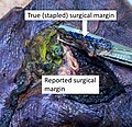
True and reported surgical margin.jpg 1,163 × 1,127; 576 KB
-
Tumors.jpg 824 × 511; 185 KB


