Category:Images of human muscles from Anatomography
Jump to navigation
Jump to search
Media in category "Images of human muscles from Anatomography"
The following 103 files are in this category, out of 103 total.
-
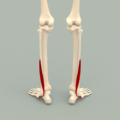
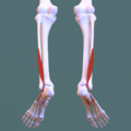
Anterior compartment of leg - Extensor digitorum longus.png 1,200 × 1,200; 975 KB
-
Anterior compartment of leg - Extensor hallucis longus.png 1,200 × 1,200; 943 KB
-
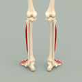
Anterior compartment of leg - Fibularis tertius.png 1,200 × 1,200; 926 KB
-
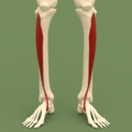
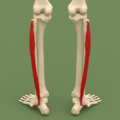
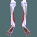
Anterior compartment of leg - Tibialis anterior.png 1,200 × 1,200; 925 KB
-
Buccinator muscle frontal.png 800 × 800; 271 KB
-
Buccinator muscle lateral.png 800 × 800; 226 KB
-
Corrugator supercilii muscle frontal.png 800 × 800; 271 KB
-
Corrugator supercilii muscle lateral.png 800 × 800; 226 KB
-
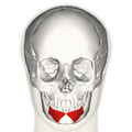
Depressor labii inferioris muscle frontal.png 800 × 800; 271 KB
-
Depressor labii inferioris muscle lateral.png 800 × 800; 228 KB
-
Depressor septi nasi muscle frontal.png 900 × 900; 322 KB
-
Depressor septi nasi muscle lateral.png 900 × 900; 270 KB
-
Extensor digitorum brevis muscle - anterior view.png 1,200 × 1,200; 993 KB
-
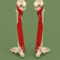
Extensor digitorum longus muscle - anteriror view.png 1,200 × 1,200; 1,021 KB
-
Extensor digitorum longus muscle - posteriror view.png 1,200 × 1,200; 980 KB
-
Extensor hallucis brevis muscle - anteriror view.png 1,200 × 1,200; 981 KB
-
Extensor hallucis longus muscle - anteriror view 2.png 1,200 × 1,200; 996 KB
-
Fascial compartments of leg (anterior compartment) - anterior view.png 1,200 × 1,200; 920 KB
-
Fascial compartments of leg (anterior compartment) - posterior view.png 1,200 × 1,200; 916 KB
-
Fascial compartments of leg (deep posterior compartment) - posterior view.png 1,200 × 1,200; 931 KB
-
Fascial compartments of leg (lateral compartment) - posterior view.png 1,200 × 1,200; 917 KB
-
Fascial compartments of leg (superficial posterior compartment) - posterior view.png 1,200 × 1,200; 910 KB
-
Fascial compartments of leg -3D.svg 1,116 × 1,203; 791 KB
-
Fibularis brevis muscle - anterior view.png 1,200 × 1,200; 986 KB
-
Fibularis brevis muscle - posterior view.png 1,200 × 1,200; 966 KB
-
Gastrocnemius muscle - posterior view.png 1,200 × 1,200; 396 KB
-
Inferior view fibularis longus muscle - anterior.png 1,200 × 1,200; 812 KB
-
Inferior view fibularis longus muscle - posterior.png 1,200 × 1,200; 843 KB
-
Inferior view of flexor digitorum longus muscle - anterior.png 1,200 × 1,200; 815 KB
-
Inferior view of flexor digitorum longus muscle - posterior.png 1,200 × 1,200; 855 KB
-
Inferior view of flexor hallucis longus muscle - anterior.png 1,200 × 1,200; 799 KB
-
Inferior view of flexor hallucis longus muscle - posterior.png 1,200 × 1,200; 837 KB
-
Inferior view of peroneus muscles - posterior.png 1,200 × 1,200; 861 KB
-
Inferior view of tibialis posterior muscle - anterior.png 1,200 × 1,200; 797 KB
-
Inferior view of tibialis posterior muscle - posterior.png 1,200 × 1,200; 832 KB
-
Lateral compartment of leg - Fibularis brevis.png 1,200 × 1,200; 924 KB
-
Lateral compartment of leg - Fibularis longus.png 1,200 × 1,200; 955 KB
-
Lateral compartment of leg.svg 1,600 × 1,600; 1.27 MB
-
Masseter muscle - anterior view - superficial part.png 640 × 640; 112 KB
-
Masseter muscle - anterior view.png 640 × 640; 115 KB
-
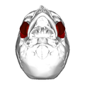
Masseter muscle - inferior view - deep part.png 640 × 640; 126 KB
-
Masseter muscle - inferior view - superficial part.png 640 × 640; 127 KB
-
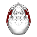
Masseter muscle - inferior view.png 640 × 640; 127 KB
-
Masseter muscle - lateral view - deep part.png 640 × 640; 98 KB
-
Masseter muscle - lateral view - superficial part.png 640 × 640; 94 KB
-
Masseter muscle - lateral view.png 640 × 640; 100 KB
-
Masseter muscle - posterior view - deep part.png 640 × 640; 91 KB
-
Masseter muscle - posterior view - superficial part.png 640 × 640; 88 KB
-
Masseter muscle - posterior view.png 640 × 640; 92 KB
-
Mentalis frontal.png 900 × 900; 323 KB
-
Mentalis lateral.png 900 × 900; 268 KB
-
Nasalis muscle frontal.png 900 × 900; 322 KB
-
Nasalis muscle lateral.png 900 × 900; 269 KB
-
Occipital bone 090 000.png 600 × 600; 114 KB
-
Occipitalis muscle back.png 1,139 × 869; 264 KB
-
Occipitalis muscle lateral.png 1,139 × 912; 288 KB
-
Peroneus muscles.svg 1,600 × 1,600; 1.35 MB
-
Peroneus tertius muscle - anteriror view.png 1,200 × 1,200; 987 KB
-
Peroneus tertius muscle - posteriror view.png 1,200 × 1,200; 961 KB
-
Plantaris muscle - anterior view.png 1,200 × 1,200; 451 KB
-
Plantaris muscle - posterior view.png 1,200 × 1,200; 436 KB
-
Popliteus muscle - posterior view.png 1,200 × 1,200; 395 KB
-
Posterior compartment of leg - flexor digitorum longus.png 1,200 × 1,200; 968 KB
-
Posterior compartment of leg - flexor hallucis longus.png 1,200 × 1,200; 944 KB
-
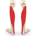
Posterior compartment of leg - gastrocnemius.png 1,200 × 1,200; 958 KB
-
Posterior compartment of leg - plantaris.png 1,200 × 1,200; 965 KB
-
Posterior compartment of leg - popliteus muscle.png 1,200 × 1,200; 934 KB
-
Posterior compartment of leg - soleus.png 1,200 × 1,200; 947 KB
-
Posterior compartment of leg - tibialis posterior.png 1,200 × 1,200; 939 KB
-
Procerus muscle frontal.png 800 × 800; 276 KB
-
Procerus muscle lateral.png 800 × 800; 227 KB
-
Soleus muscle - anterior view.png 1,200 × 1,200; 428 KB
-
Soleus muscle - posterior view.png 1,200 × 1,200; 510 KB
-
Splenius cervicis muscle back.png 900 × 900; 239 KB
-
Splenius cervicis muscle frontal.png 900 × 900; 262 KB
-
Splenius cervicis muscle lateral.png 900 × 900; 191 KB
-
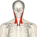
Sternomastoid muscle back.png 900 × 900; 243 KB
-
Sternomastoid muscle back2.png 900 × 900; 413 KB
-
Sternomastoid muscle frontal.png 900 × 900; 273 KB
-
Sternomastoid muscle frontal2.png 900 × 900; 467 KB
-
Sternomastoid muscle lateral.png 900 × 900; 200 KB
-
Sternomastoid muscle lateral2.png 900 × 900; 343 KB
-
Superior view fibularis longus muscle - posterior.png 1,200 × 1,200; 985 KB
-
Superior view of flexor digitorum longus muscle - anterior.png 1,200 × 1,200; 1,003 KB
-
Superior view of flexor hallucis longus muscle - posterior.png 1,200 × 1,200; 970 KB
-
Superior view of peroneus muscles - anterior.png 1,200 × 1,200; 1,020 KB
-
Superior view of peroneus muscles - posterior.png 1,200 × 1,200; 993 KB
-
Superior view of tibialis posterior muscle - anterior.png 1,200 × 1,200; 999 KB
-
Superior view of tibialis posterior muscle - posterior.png 1,200 × 1,200; 971 KB
-
Temporal muscle - anterior view.png 640 × 640; 201 KB
-
Temporal muscle - inferior view.png 640 × 640; 138 KB
-
Temporal muscle - lateral view.png 640 × 640; 237 KB
-
Temporal muscle - posterior view.png 640 × 640; 152 KB
-
Temporal muscle - superior view.png 640 × 640; 71 KB
-
Temporoparietalis muscle frontal.png 1,139 × 869; 335 KB
-
Temporoparietalis muscle lateral.png 1,139 × 912; 348 KB
-
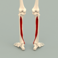
Tibialis anterior muscle - anteriror view.png 1,200 × 1,200; 986 KB
-
Tibialis anterior muscle - posteriror view.png 1,200 × 1,200; 977 KB
-
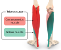
Triceps surae.svg 1,365 × 1,139; 650 KB
-
Zygomaticus major muscle frontal.png 600 × 600; 178 KB
-
Zygomaticus major muscle lateral.png 600 × 600; 152 KB
-
Zygomaticus minor muscle frontal.png 900 × 900; 322 KB
-
Zygomaticus minor muscle lateral.png 900 × 900; 271 KB


