Category:Human metatarsus
Jump to navigation
Jump to search
Subcategories
This category has the following 9 subcategories, out of 9 total.
- SVG human metatarsus (3 F)
2
- Second metatarsal bone (11 F)
3
- Third metatarsal bone (13 F)
4
- Fourth metatarsal bone (13 F)
5
F
- Human first metatarsal bone (25 F)
M
- Metatarsal accessory bones (16 F)
Media in category "Human metatarsus"
The following 104 files are in this category, out of 104 total.
-
3D CT Reconstruction of Distal tibia fracture.gif 988 × 918; 9.32 MB
-
Calcaneus Fracture.jpg 2,270 × 1,542; 256 KB
-
Calcar Calcanei 01.jpg 2,270 × 1,604; 1.27 MB
-
Calcar Calcanei 02.jpg 2,272 × 1,704; 1.75 MB
-
CT 3D human Foot Skin and Bone.jpeg 400 × 400; 17 KB
-
Cunningham’s Text-book of Anatomy (1914) - Fig 256.png 1,732 × 2,106; 2.21 MB
-
Dixon's Manual of human osteology (1912) - Fig 094.png 1,382 × 1,080; 446 KB
-
Dixon's Manual of human osteology (1912) - Fig 095.png 1,502 × 1,094; 481 KB
-
Flickr - BioDivLibrary - n296 w1150 (3).jpg 1,236 × 862; 140 KB
-
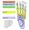
Foot bones - metatarsus and phalanges.jpg 960 × 720; 96 KB
-
Foot bones - tarsus, metatarsus-zh.jpg 960 × 720; 175 KB
-
Foot bones - tarsus, metatarsus.jpg 960 × 720; 99 KB
-
Foot bones.jpg 657 × 1,305; 85 KB
-
Foot retro.JPG 2,288 × 1,712; 1.35 MB
-
Foot.png 214 × 356; 10 KB
-
Footx.jpg 619 × 456; 34 KB
-
Fussgelenke.jpg 1,386 × 733; 155 KB
-
Gray foot bone lateral view.gif 500 × 179; 23 KB
-
Gray foot bone medial view.gif 500 × 189; 19 KB
-
Gray268 - Mratatarsus.png 638 × 1,195; 395 KB
-
Gray269 - Mratatarsus.png 649 × 1,184; 354 KB
-
Gray284.png 321 × 450; 32 KB
-
Gray285.png 289 × 450; 26 KB
-
Gray286.png 290 × 400; 19 KB
-
Gray287.png 300 × 400; 21 KB
-
Gray288 he.png 267 × 375; 18 KB
-
Gray288.png 267 × 375; 19 KB
-
Gray289.png 492 × 600; 27 KB
-
Gray290 - Mratatarsus.png 500 × 189; 85 KB
-
Gray291 - Metatarsus.png 500 × 179; 84 KB
-
Gray354.png 550 × 424; 39 KB
-
Gray355.png 600 × 487; 57 KB
-
Gray358 int.png 621 × 700; 172 KB
-
Gray358.png 621 × 700; 67 KB
-
Gray360.png 444 × 550; 58 KB
-
Hallux varus congenitus.jpg 436 × 704; 32 KB
-
Homo luzonensis metatarsal.jpg 1,595 × 1,230; 69 KB
-
Human right foot bones 3D print.jpg 790 × 647; 313 KB
-
JonesFracture1.png 458 × 899; 234 KB
-
Left Metatarsal bones - animation01.gif 450 × 450; 1.42 MB
-
Left Metatarsal bones - animation02.gif 450 × 450; 1.38 MB
-
Left Metatarsal bones01 anterior view.png 4,500 × 4,500; 1.95 MB
-
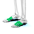
Left Metatarsal bones02 lateral view.png 4,500 × 4,500; 2.02 MB
-
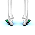
Left Metatarsal bones03 posterior view.png 4,500 × 4,500; 1.83 MB
-
Left Metatarsal bones04 medial view.png 4,500 × 4,500; 1.99 MB
-
Left Metatarsal bones05 anterior view.png 4,500 × 4,500; 1.24 MB
-
Left Metatarsal bones06 lateral view.png 4,500 × 4,500; 1.12 MB
-
Left Metatarsal bones07 posterior view.png 4,500 × 4,500; 1.32 MB
-
Left Metatarsal bones08 medial view.png 4,500 × 4,500; 1.2 MB
-
Medical X-Ray imaging BZB03 nevit.jpg 2,486 × 1,974; 372 KB
-
Medical X-Ray imaging DAB03 nevit.jpg 1,784 × 2,384; 206 KB
-
Medical X-Ray imaging IIQ05 nevit.jpg 2,384 × 1,776; 1.76 MB
-
Medical X-Ray imaging KVW05 nevit.jpg 2,384 × 1,784; 1.51 MB
-
Medical X-Ray imaging LOL05 nevit.jpg 1,184 × 1,736; 813 KB
-
Medical X-Ray imaging LOM05 nevit.jpg 1,288 × 936; 556 KB
-
Medical X-Ray imaging NBE06 nevit.jpg 2,384 × 1,784; 283 KB
-
Metatarsal bones - animation01.gif 450 × 450; 1.94 MB
-
Metatarsal bones01 - superior view.png 4,500 × 4,500; 1.83 MB
-
Metatarsal bones02 - inferior view.png 4,500 × 4,500; 2.07 MB
-
Metatarsal bones03.png 4,500 × 4,500; 3.51 MB
-
Metatarsal bones04.png 4,500 × 4,500; 3.24 MB
-
Metatarsal bones05.png 4,500 × 4,500; 3.27 MB
-
Metatarsal bones06.png 4,500 × 4,500; 3.14 MB
-
Metatarsal bones07.png 4,500 × 4,500; 1.78 MB
-
Metatarsal bones08.png 4,500 × 4,500; 1.69 MB
-
Metatarsal bones09.png 4,500 × 4,500; 1.48 MB
-
Metatarsal pseudo-epiphysis.jpg 916 × 1,164; 232 KB
-
Metatarsal pseudo-epiphysis.svg 458 × 582; 241 KB
-
Metatarsus.jpg 960 × 720; 74 KB
-
Nitti MetatarsalGuard.jpg 1,314 × 949; 521 KB
-
Ospied-en.svg 379 × 395; 53 KB
-
Ospied-la.svg 378 × 379; 53 KB
-
Polydactyly 01 Lfoot AP.jpg 717 × 1,452; 153 KB
-
Polydactyly 01 Rfoot AP.jpg 747 × 1,479; 151 KB
-
Scaphocephaly feet.jpg 1,911 × 1,457; 931 KB
-
Sesamoidbone.png 1,376 × 880; 409 KB
-
SesamoidBonesOfFoot.svg 1,362 × 1,099; 388 KB
-
Slide1xzxz-ar.jpg 960 × 720; 134 KB
-
Slide1xzxz.JPG 960 × 720; 73 KB
-
Slide2dede.JPG 960 × 720; 54 KB
-
Slide2xzxzx zh.jpg 960 × 720; 106 KB
-
Slide2xzxzx-ar.jpg 960 × 720; 120 KB
-
Slide2xzxzx.JPG 960 × 720; 71 KB
-
Slide3CEC2-ar.jpg 960 × 720; 169 KB
-
Slide3CEC2.JPG 960 × 720; 76 KB
-
Slide5ecce-ar.jpg 960 × 720; 132 KB
-
Slide5ecce.JPG 960 × 720; 71 KB
-
Slide6CEC5.JPG 960 × 720; 87 KB
-
Slide7CEC6.JPG 960 × 720; 76 KB
-
Sobo 1909 228.png 2,799 × 1,590; 12.75 MB
-
Sobo 1909 313.png 1,040 × 1,660; 4.95 MB
-
Sobo 1909 314.png 1,084 × 1,656; 5.15 MB
-
Sobo 1909 315.png 2,504 × 1,720; 12.34 MB
-
Tape21.png 379 × 487; 250 KB
-
Tape22.png 758 × 487; 404 KB
-
Tape23.png 758 × 487; 416 KB
-
Testut's Treatise on Human Anatomy (1911) - Vol 1 - Fig 375.png 1,578 × 2,134; 3.15 MB
-
William Cheselden feet.jpg 844 × 1,323; 285 KB
-
X-ray.jpg 1,036 × 775; 87 KB
-
Zehenknochen-xrayS0 I0.JPG 1,050 × 844; 100 KB


