Category:Histopathology of melanoma
Jump to navigation
Jump to search
Subcategories
This category has the following 17 subcategories, out of 17 total.
A
L
M
N
O
P
Media in category "Histopathology of melanoma"
The following 84 files are in this category, out of 84 total.
-
 BRAF V600E mutant melanoma.jpg 2,080 × 1,542; 746 KB
-
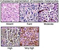 Cytoplasmic pigmentation of neoplastic melanocytes.jpg 668 × 565; 146 KB
-
 Grey horse melanoma 1.JPG 2,048 × 1,536; 3.24 MB
-
 Histopathology of a biopsy of a melanoma metastasis to the brain.jpg 2,048 × 1,532; 470 KB
-
 Histopathology of invasive melanoma, high magnification.jpg 2,048 × 1,532; 412 KB
-
 Histopathology of Malignant melanoma.jpg 590 × 502; 175 KB
-
 Histopathology of melanoma, HE.jpg 454 × 710; 167 KB
-
 Histopathology of melanoma.jpg 979 × 711; 343 KB
-
 Lymph node with almost complete replacement by metastatic melanoma.jpg 12,991 × 8,609; 41.39 MB
-
 Lymph node with metastatic melanoma - by Gabriel Caponetti, MD.jpg 12,641 × 8,291; 91.52 MB
-
 Lymph node with metastatic melanoma.jpg 2,598 × 1,722; 5.32 MB
-
 Malignant melanoma (1) at thigh Case 01.jpg 600 × 452; 72 KB
-
 Malignant melanoma (3) at thigh Case 01.jpg 600 × 452; 82 KB
-
 Malignant melanoma (4) at thigh Case 01.jpg 600 × 452; 72 KB
-
 Malignant melanoma cns.jpg 2,080 × 1,542; 1.01 MB
-
 Malignant melanoma HMB45.jpg 2,080 × 1,542; 991 KB
-
 Malignant melanoma in situ - alt -- very high mag.jpg 4,272 × 2,848; 4.2 MB
-
 Malignant melanoma in situ -- high mag.jpg 4,272 × 2,848; 5.21 MB
-
 Malignant melanoma in situ -- intermed mag.jpg 4,272 × 2,848; 4.8 MB
-
 Malignant melanoma in situ -- low mag.jpg 4,272 × 2,848; 4.99 MB
-
 Malignant melanoma in situ -- very high mag.jpg 2,848 × 4,272; 4.3 MB
-
 Malignant melanoma in situ -- very low mag.jpg 4,272 × 2,848; 4.84 MB
-
 Melanoma (6).jpg 2,331 × 1,800; 2.04 MB
-
 Melanoma - cytology field stain.jpg 3,324 × 2,348; 3.05 MB
-
 Melanoma 03.jpg 4,080 × 3,072; 4.58 MB
-
 Melanoma cell shapes.jpg 467 × 564; 120 KB
-
 Melanoma CNS met smear HE.jpg 2,080 × 1,542; 782 KB
-
 Melanoma metastasis in heart.jpg 802 × 830; 112 KB
-
 Melanoma metastasis in hepar.jpg 1,122 × 1,122; 470 KB
-
 Melanoma mimicking CC.jpg 2,080 × 1,542; 841 KB
-
 Melanoma resembling a squamous cell carcinoma.jpg 2,048 × 1,532; 820 KB
-
 Melanoma40x.JPG 2,048 × 1,536; 1.2 MB
-
 Mus musculus melanoma skin cancer cells in UFV.jpg 3,456 × 2,304; 5.13 MB
-
 Nesting of intraepidermal melanocytes.jpg 675 × 569; 162 KB
-
 Neuropathology case VII 01.jpg 2,080 × 1,542; 819 KB
-
 Neuropathology case VII 02.jpg 2,080 × 1,542; 741 KB
-
 Neuropathology case VII 03.jpg 2,080 × 1,542; 831 KB
-
 Neuropathology case VII 04.jpg 2,080 × 1,542; 620 KB
-
 Neuropathology case VII 05.jpg 2,080 × 1,542; 1.14 MB
-
 Neuropathology case VII 06.jpg 2,080 × 1,542; 685 KB
-
 Nodular melanoma with heavy melanin pigmentation.jpg 693 × 520; 161 KB
-
 Nodular melanoma, high magnification.jpg 693 × 520; 130 KB
-
 Nodular melanoma, medium magnification.jpg 693 × 520; 201 KB
-
 Retinal melanoma (low zoom).jpg 1,206 × 887; 230 KB
-
 Retinal melanoma.jpg 1,188 × 996; 237 KB
-
 Scatter of intraepidermal melanocytes.jpg 659 × 575; 175 KB
-
 Sentinel lymph node positive for melanoma micrometastases.png 320 × 253; 220 KB
-
 Skin MMIS HP 14PY***.jpg 2,048 × 1,536; 1.69 MB
-
 Skin Tumors-008.jpg 1,280 × 1,024; 786 KB
-
 Skin Tumors-042.jpg 1,280 × 1,024; 821 KB
-
 Skin Tumors-043.jpg 1,280 × 1,024; 292 KB
-
 Skin Tumors-055.jpg 1,280 × 1,024; 895 KB
-
 Skin Tumors-056.jpg 1,280 × 1,024; 815 KB
-
 Skin Tumors-057.jpg 1,280 × 1,024; 785 KB
-
 Skin Tumors-062.jpg 1,280 × 1,024; 288 KB
-
 Skin Tumors-068.jpg 1,280 × 1,024; 259 KB
-
 Skin Tumors-070.jpg 1,280 × 1,024; 731 KB
-
 Skin Tumors-077.jpg 1,280 × 1,024; 871 KB
-
 Skin Tumors-079.jpg 1,280 × 1,024; 287 KB
-
 Skin Tumors-343.jpg 1,280 × 1,024; 273 KB
-
 Skin Tumors-361.jpg 1,280 × 1,024; 256 KB
-
 Skin Tumors-368.jpg 1,280 × 1,024; 1.04 MB
-
 Skin Tumors-372.jpg 1,280 × 1,024; 787 KB
-
 Skin Tumors-378.jpg 1,280 × 1,024; 948 KB
-
 Skin Tumors-613.jpg 1,280 × 1,024; 983 KB
-
 Skin Tumors-615.jpg 1,280 × 1,024; 871 KB
-
 Skin Tumors-616.jpg 1,280 × 1,024; 746 KB
-
 Skin Tumors-647.jpg 1,280 × 1,024; 1.04 MB
-
 Skin Tumors-P6231209.jpg 1,600 × 1,200; 1.42 MB
-
 Skin Tumors-P6231210.jpg 1,600 × 1,200; 1.42 MB
-
 Skin Tumors-P6231217.jpg 1,600 × 1,200; 1.53 MB
-
 Skin Tumors-P6231218.jpg 1,600 × 1,200; 1.41 MB
-
 Skin Tumors-P6231222.jpg 1,600 × 1,200; 1.32 MB
-
 Skin Tumors-P6231223.jpg 1,600 × 1,200; 1.25 MB
-
 Skin Tumors-P6231228.jpg 1,600 × 1,200; 1.53 MB
-
 Skin Tumors-P6231231.jpg 1,600 × 1,200; 1.37 MB
-
 Skin Tumors-P6231232.jpg 1,600 × 1,200; 1.4 MB
-
 Skin Tumors-P6231244.jpg 1,600 × 1,200; 1.44 MB
-
 Skin Tumors-P6231245.jpg 1,600 × 1,200; 1.41 MB
-
 Skin Tumors-P6231250.jpg 1,600 × 1,200; 1.69 MB
-
 Skin Tumors-P6231252.jpg 1,600 × 1,200; 1.24 MB
-
 Skin Tumors-P6241254.jpg 1,600 × 1,200; 1.6 MB
-
 Skin Tumors-P6241255.jpg 1,600 × 1,200; 1.53 MB
-
 Superficially spreading melanoma.jpg 693 × 520; 132 KB


_at_thigh_Case_01.jpg)
_at_thigh_Case_01.jpg)
_at_thigh_Case_01.jpg)


.jpg)


.jpg)


