Category:Computed tomography images
Jump to navigation
Jump to search
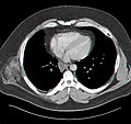
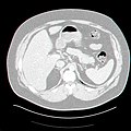
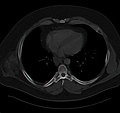
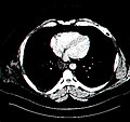
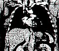
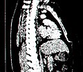
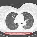
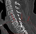
English: Images made with computed tomography (CT)
Subcategories
This category has the following 14 subcategories, out of 14 total.
Media in category "Computed tomography images"
The following 135 files are in this category, out of 135 total.
-
9 volt battery microCT 1272.jpg 5,121 × 7,057; 10.75 MB
-
Abdomen-Window(L0)(W350)Axial.jpg 468 × 442; 59 KB
-
Abdomen-Window(L0)(W350)Coronal.jpg 468 × 406; 71 KB
-
Abdomen-Window(L0)(W350)Sagittal.jpg 468 × 406; 62 KB
-
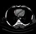
Axial plane CT scan of the thorax illustrative image.jpg 1,163 × 493; 359 KB
-
Axial-Abdomen(L-200)(W600).jpg 512 × 512; 70 KB
-
Axial-Abdomen(L0)(W1000).jpg 512 × 512; 54 KB
-
Axial-Abdomen(L0)(W200).jpg 512 × 512; 100 KB
-
Axial-Abdomen(L0)(W600).jpg 512 × 512; 70 KB
-
Axial-Abdomen(L200)(W600).jpg 512 × 512; 59 KB
-
Back-Projection reconstructions of Shepp-Logan phantom at different angles.png 1,920 × 963; 214 KB
-
Blausen 0222 CervicalSpine.png 768 × 1,024; 653 KB
-
Blausen 0223 CesareanDelivery.png 1,600 × 1,200; 915 KB
-
Bone-Window(L600)(W1600)Axial.jpg 468 × 442; 27 KB
-
Bone-Window(L600)(W1600)Coronal.jpg 468 × 406; 32 KB
-
Bone-Window(L600)(W1600)Sagittal.jpg 468 × 406; 31 KB
-
Brain-isolines.png 641 × 565; 101 KB
-
Brain-Window(L35)(W70)Axial.jpg 468 × 442; 55 KB
-
Brain-Window(L35)(W70)Coronal.jpg 468 × 406; 94 KB
-
Brain-Window(L35)(W70)Sagittal.jpg 468 × 406; 69 KB
-
Cerebral Folate Deficiency - Cerebral CT-scan at 4 years old.png 2,986 × 1,556; 644 KB
-
Cochlear aplasia with dysplastic vestibule.jpg 504 × 332; 101 KB
-
Computed tomography 2.png 1,280 × 853; 2.07 MB
-
Computed Tomography slice of the lungs with normal processing.jpg 515 × 515; 131 KB
-
Computed Tomography slice of the thorax with normal processing.jpg 515 × 515; 110 KB
-
Computed tomography.jpg 1,280 × 853; 180 KB
-
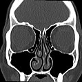
Coronal plane CT scan of the paranasal sinuses illustrative image.jpg 1,016 × 508; 153 KB
-
CT and MRI of a red-eared slider (Trachemys scripta) - journal.pone.0017879.g001.png 2,009 × 2,941; 2.46 MB
-
CT detector artifact.jpg 1,024 × 1,024; 215 KB
-
CT EIT Superposition.jpg 512 × 489; 127 KB
-
CT scan and MRI brain image of basal ganglia at CKD and AKI patient.png 1,181 × 1,852; 673 KB
-
CT Scan General Illustration Upper Abdomen.jpg 892 × 831; 202 KB
-
CT Scan General Illustration.jpg 907 × 895; 539 KB
-
CT Scan Liver-Kidneys.jpg 5,017 × 3,345; 1.54 MB
-
CT scan of a patient with Descending Necrotizing Mediastinitis.jpg 468 × 235; 37 KB
-
CT Scan Thorax Air -1000 HU Window Level.jpg 917 × 895; 642 KB
-
CT Scan Thorax Liver 60 HU Window Level.jpg 917 × 895; 217 KB
-
CT Scan Thorax Liver.jpg 917 × 895; 193 KB
-
CT Scan Thorax Lung -700 HU Window Level.jpg 917 × 895; 188 KB
-
CT Scan Thorax Water 0 HU Window Level.jpg 917 × 895; 370 KB
-
CT scans (5940498869).jpg 410 × 362; 184 KB
-
CT scans (5941057880).jpg 817 × 598; 598 KB
-
CT ScoutView.jpg 880 × 1,024; 122 KB
-
CT tooth artifact.jpg 1,024 × 1,024; 284 KB
-
CT UNILAB 04.png 174 × 364; 61 KB
-
CT UNILAB 05.png 137 × 364; 56 KB
-
CT Unilab 06.png 115 × 151; 29 KB
-
CT Unilab 08.png 141 × 173; 22 KB
-
CT Unilab 09.png 263 × 169; 50 KB
-
CT-Artefakt-Becken-Metall.jpg 886 × 831; 70 KB
-
CT-Artefakt-HWS-BWS-sagittal.jpg 679 × 646; 40 KB
-
CT-PET.jpg 516 × 395; 41 KB
-
CT-Standard-Dose-2.50-Lung-Calcified-Nodule.jpg 1,920 × 1,080; 579 KB
-
Ct-workstation-neck.jpg 1,026 × 1,026; 225 KB
-
CT. Glenoidal retroversion of around 5°..jpg 798 × 1,120; 335 KB
-
CT. Glenoidal retroversion..jpg 798 × 560; 74 KB
-
CTAbdoN2008.jpg 3,072 × 2,304; 533 KB
-
CtFluoroImages.jpg 955 × 439; 179 KB
-
CTscanportalveinandliver.jpg 510 × 466; 35 KB
-
DeepLearningReconstruction.png 2,502 × 600; 617 KB
-
Doctor and patient looking at CT Scan of Lungs.jpg 6,000 × 4,000; 6.31 MB
-
E cig tomography of chests mm6836e1-F1.gif 308 × 900; 73 KB
-
Exceptional preservation of nerves, digestive tract and stomachal content.png 2,517 × 3,275; 4.28 MB
-
Facets normal ct w text better contrast.jpg 600 × 585; 139 KB
-
Foam ball.png 1,101 × 1,011; 971 KB
-
Fossilized Mosasaur Tooth.png 2,065 × 1,472; 1.36 MB
-
Gallstone µCT.jpg 1,820 × 7,251; 4.34 MB
-
Geometrik Morfometrik.jpg 640 × 280; 47 KB
-
Haller cell ct.JPG 377 × 379; 19 KB
-
Halloweensign.jpg 1,536 × 2,048; 941 KB
-
HiatushernieMPR.png 1,534 × 955; 1.11 MB
-
Horizontal Impaction.jpg 3,298 × 2,638; 2.43 MB
-
Image-Infiltration-C5-C6-Fluro-Scanner.jpg 1,680 × 1,680; 181 KB
-
Left Maxillary Sinusitis on CT Slice.jpg 942 × 592; 70 KB
-
Lenkgehaeuse 3D-CT.png 815 × 472; 389 KB
-
Liver-Window(L100)(W200)Axial.jpg 468 × 442; 41 KB
-
Liver-Window(L100)(W200)Coronal.jpg 468 × 406; 67 KB
-
Liver-Window(L100)(W200)Sagittal.jpg 468 × 406; 56 KB
-
Lung-Window(L-600)(W1600)Axial.jpg 468 × 442; 40 KB
-
Lung-Window(L-600)(W1600)Coronal.jpg 468 × 406; 39 KB
-
Lung-Window(L-600)(W1600)Sagittal.jpg 468 × 406; 35 KB
-
MBq digital-radiograph.jpg 1,207 × 403; 65 KB
-
Micro-CT braided polymer rope 2D lateral view 2.jpg 2,123 × 1,437; 2.89 MB
-
Micro-CT braided polymer rope 2D lateral view.jpg 2,123 × 1,437; 2.25 MB
-
Micro-CT braided polymer rope 2D top view zoom.jpg 2,123 × 1,451; 2.23 MB
-
Micro-CT braided polymer rope 2D top view.jpg 2,123 × 1,451; 1.19 MB
-
Micro-CT braided polymer rope 3D 01.jpg 2,139 × 1,451; 1.82 MB
-
Micro-CT braided polymer rope 3D 02.jpg 2,139 × 1,451; 1.92 MB
-
Micro-CT braided polymer rope 3D 03.jpg 2,139 × 1,451; 1.67 MB
-
Micro-CT braided polymer rope 3D 04.jpg 2,139 × 1,451; 2.54 MB
-
Micro-CT braided polymer rope 3D 05.jpg 2,139 × 1,451; 2.13 MB
-
Micro-CT braided polymer rope 3D 06.jpg 2,139 × 1,451; 1.69 MB
-
Micro-CT braided polymer rope 3D 07.jpg 2,139 × 1,451; 2.02 MB
-
Micro-CT braided polymer rope 3D 08.jpg 2,139 × 1,451; 2.37 MB
-
Micro-CT braided polymer rope 3D 09.jpg 2,139 × 1,451; 1.68 MB
-
Micro-CT braided polymer rope 3D 10.jpg 2,139 × 1,451; 1.76 MB
-
Mosasaur Tooth.jpg 2,065 × 1,472; 837 KB
-
Mpr and mip sts.png 1,024 × 1,024; 1,018 KB
-
Mpr and sts.jpg 1,024 × 1,024; 452 KB
-
Nautilus Section cut Logarithmic spiral.jpg 972 × 377; 121 KB
-
Oncology radiation therapy planning.jpg 3,264 × 2,448; 1.75 MB
-
Optimising CT Image Analysis & Visualisation For the Clinical Environment.pdf 1,239 × 1,754, 61 pages; 1.25 MB
-
Orbita Cristalino.jpg 455 × 387; 46 KB
-
Part to CAD Analysis - 2.jpg 160 × 131; 4 KB
-
Part to CAD Analysis - 2.png 544 × 332; 133 KB
-
Part to Part Analysis - 2.png 160 × 98; 27 KB
-
PF-Window(L35)(W150)Axial.jpg 468 × 442; 53 KB
-
PF-Window(L35)(W150)Coronal.jpg 468 × 406; 83 KB
-
PF-Window(L35)(W150)Sagittal.jpg 468 × 406; 64 KB
-
PikiWiki Israel 15787 Subtle diagnosis.jpg 512 × 758; 62 KB
-
PikiWiki Israel 15788 Subtle diagnosis.jpg 1,711 × 1,140; 149 KB
-
PMC4441996 CRIONM2015-341064.001.png 512 × 224; 87 KB
-
Right dense MCA sign as seen on CT brain.jpg 3,456 × 4,608; 5.3 MB
-
SADDLE PE.JPG 1,053 × 876; 96 KB
-
Sagital sinus thrombus.JPG 873 × 1,058; 100 KB
-
Series 001 (3-PL LOC).png 256 × 256; 65 KB
-
Serum urate deposition.jpg 484 × 186; 19 KB
-
Spine-Window(L60)(W300)Axial.jpg 468 × 442; 52 KB
-
Spine-Window(L60)(W300)Coronal.jpg 468 × 406; 70 KB
-
Spine-Window(L60)(W300)Sagittal.jpg 468 × 406; 61 KB
-
Theragripper attachment to rat colon and retention upon rectal delivery.jpg 1,050 × 819; 279 KB
-
TORNAI-SpectrumOfMedicalImaging.jpg 720 × 504; 117 KB
-
Tracheal diverticulum annotated HD.jpg 1,920 × 1,080; 448 KB
-
Volume graphic assembly analysis.jpg 160 × 142; 11 KB
-
Volume rendered CT urography.jpg 296 × 431; 25 KB
-
Wegener's Granulomatosis - CT scan Case 190 (5958079080).jpg 512 × 440; 55 KB
-
ZProject.jpg 763 × 789; 250 KB
-
Туб3.jpg 2,848 × 3,268; 5.23 MB
-
“The Skull of You” – MicroCT scan of a mouse skull.jpg 2,400 × 2,400; 1.42 MB

