Category:Chemistry images with transparent background
Jump to navigation
Jump to search
Subcategories
This category has the following 4 subcategories, out of 4 total.
B
R
Media in category "Chemistry images with transparent background"
The following 58 files are in this category, out of 58 total.
-
(-) and (+) Phantasmidine.png 1,865 × 1,238; 103 KB
-
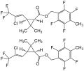
(1R,3R)-rel-Tefluthrine Formulae V.1.svg 729 × 618; 57 KB
-
(±)-Meclozine enantiomers structural formulae.png 1,656 × 2,228; 21 KB
-
1a2o structure.png 2,500 × 2,000; 1.17 MB
-
1axc tricolor.png 1,108 × 1,196; 582 KB
-
1CQ0 crystallography.png 820 × 638; 133 KB
-
1kko.png 1,000 × 1,000; 438 KB
-
1NLRribbon.png 752 × 845; 248 KB
-
1r4o.png 960 × 720; 257 KB
-
201309 DNAparts.png 400 × 400; 23 KB
-
201812 DNA double-strand A.svg 512 × 2,199; 22 KB
-
2bq0 14-3-3.png 1,447 × 755; 408 KB
-
Acethylcholinesterase TC 1EA5.png 960 × 720; 317 KB
-
ADN static.png 423 × 657; 79 KB
-
AgeingDw001.png 2,173 × 1,694; 530 KB
-
Alpha-sodium-metavanadate-chain-from-xtal-1974-CM-3D-polyhedra.png 2,150 × 739; 219 KB
-
Ammonium-metavanadate-3D-polyhedra.png 1,100 × 804; 218 KB
-
Amyloid beta fibrils.png 745 × 628; 301 KB
-
Beryllium-hydride-3D-polyhedra-A.png 1,100 × 939; 255 KB
-
Beta-sodium-metavanadate-chain-from-xtal-1984-CM-3D-polyhedra.png 2,150 × 723; 333 KB
-
Butyrylcholinesterase 1P0I.png 1,000 × 915; 540 KB
-
Cas9 5AXW plain.png 2,245 × 1,651; 3.56 MB
-
Cellulase 1JS4.png 2,160 × 960; 1.17 MB
-
Cryo-EM structure of the human ether-a-go-go related K+ channel.png 640 × 480; 200 KB
-
Crystal structure of modified Gramicidin S horizontally.png 571 × 294; 80 KB
-
Crystal Structure of the Human vaccinia-related kinase.png 640 × 480; 119 KB
-
Docking toxapy.png 451 × 487; 321 KB
-
Dodecahedrane.svg 405 × 420; 7 KB
-
Ghrelin-3D-predicted.png 1,500 × 1,500; 438 KB
-
GLP-1R complex.png 2,160 × 3,840; 2.06 MB
-
Golimumab 5yoy.png 640 × 480; 93 KB
-
Hemoglobin E (Glu26Lys) 1yvt.png 640 × 480; 298 KB
-
HRP-xray.png 1,300 × 1,584; 938 KB
-
Human cystic fibrosis transmembrane conductance regulator (CFTR).png 640 × 480; 234 KB
-
Hydrogen-chloride-elpot-transparent-3D-balls.png 1,100 × 936; 180 KB
-
Koffein-1.png 204 × 190; 22 KB
-
KRAS protein 3GFT.png 1,035 × 859; 526 KB
-
Liraglutide cartoon 4APD.png 2,560 × 954; 156 KB
-
Ossirano struttura modello.png 219 × 108; 9 KB
-
Pasta domain from 3m9g.png 640 × 480; 100 KB
-
PDB 2GUO - MHC HLA-A2 in complex with Melan-A-MART-1 27-35 peptide.png 970 × 1,243; 479 KB
-
PDB 2jr8 EBI.png 800 × 600; 118 KB
-
Preproghrelin 1P7X.png 1,567 × 1,441; 383 KB
-
Protein CECR1 PDB 3LGD.png 1,200 × 1,000; 472 KB
-
Protein CHEK2 PDB 1gxc.png 961 × 735; 375 KB
-
Protein MAX PDB 1an2.png 721 × 559; 99 KB
-
Protein MSH2 PDB 2o8b.png 924 × 731; 653 KB
-
Protein PMS2 PDB 1ea6.png 1,151 × 731; 444 KB
-
Protein PRPF40A PDB 1uzc.png 853 × 509; 165 KB
-
Rhodium-trichloride-layer-from-xtal-1964-3D-polyhedra.png 1,100 × 792; 164 KB
-
Rhodium-trichloride-layers-xtal-1964-3D-polyhedra-B.png 1,100 × 917; 130 KB
-
Rhodium-trichloride-layers-xtal-1964-3D-polyhedra.png 1,100 × 957; 250 KB
-
Rigid rotor HCl model (2).png 1,379 × 749; 38 KB
-
Structure of a bacterial cellulose synthase.png 929 × 785; 308 KB
-
Thallium(I)-iodide-Cmcm-Tl-coord-CM-3D-polyhedra.png 1,027 × 1,100; 272 KB
-
Tin-dichloride-elpot-transparent-3D-balls.png 900 × 710; 228 KB
-
Unwound DNA Duplex.png 2,000 × 4,032; 521 KB
-
Ácido desoxirribonucleico (DNA).png 1,280 × 720; 720 KB


